







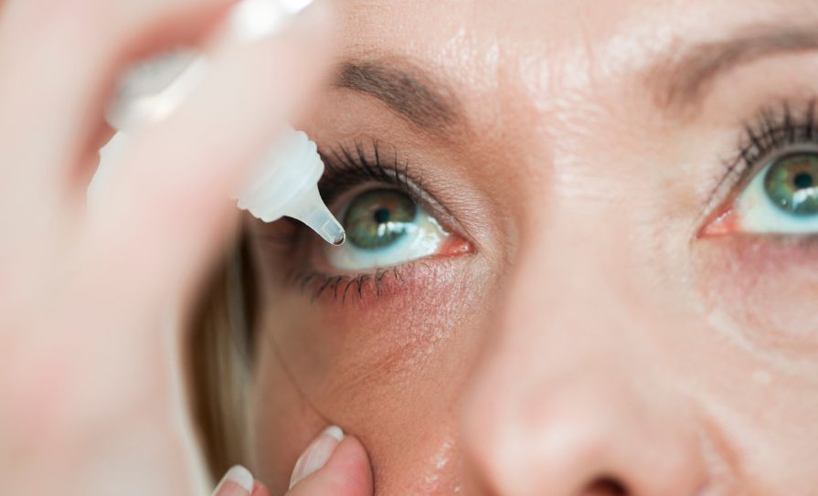
Preoperative ocular surface disease should be identified and optimised before cataract and refractive surgery
Keratoconjunctivitis sicca, or dry eye disease (DED), is defined as a multifactorial disorder of tear film homeostasis and ocular symptoms, marked by tear film instability, hyperosmolarity, ocular surface inflammation, or neurosensory abnormalities.
DED affects around one-third of the UK population and its impact on quality-of-life is frequently underestimated.
Individuals who suffer from DED may experience ocular discomfort: Itching, dryness, burning, or a foreign body sensation on the surface of the eye.
Beyond the symptoms of physical irritation, it can also affect visual function, with reports of blurred vision, fluctuations in vision when blinking, and eye fatigue. The impact of DED on a patient’s quality-of-life can be substantial, affecting both physical and mental health.
Patients suffering from DED show an increased risk of unemployment and absenteeism when compared to those without, and a systematic review and meta-analysis found that depression and anxiety were more prevalent and more severe among patients with DED. While it is frequently framed as a chronic quality-of-life condition, this article focuses specifically on its relevance to the surgical patient, examining how and why preoperative ocular surface disease should be identified and optimised before cataract and refractive surgery.
Characteristics
At its core, DED is a disorder of tear film homeostasis, in which the equilibrium between tear production, distribution, and evaporation is disrupted. This imbalance leads to instability of the tear film and, over time, to inflammation of the ocular surface, the mechanism underlying the burden described above. Appreciating how this system functions and where it tends to fail is essential to understanding both how the disease presents and how it should be assessed.
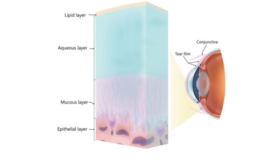
The tear film consists of three separate layers: The thinnest, outermost lipid layer is secreted by the meibomian glands. The primary function of this layer is to delay the evaporation of the aqueous layer beneath it.
The aqueous layer is the middle and most substantial layer, produced by the main lacrimal gland as well as the accessory glands of Krause and Wolfring, located within the conjunctiva. It maintains ocular-surface hydration, nourishes the avascular cornea, helps to clear debris, and provides antimicrobial defence. The innermost layer – the mucin layer – is primarily produced by the conjunctival goblet cells. It anchors the tear film to the epithelium and facilitates its even distribution across the otherwise hydrophobic epithelial surface. Beyond its role in surface protection and lubrication, the tear film also functions as the eye’s outermost refractive surface, meaning any instability or irregularity directly influences the quality of the optical image reaching the cornea. This becomes clinically significant in the context of preoperative assessment, particularly in cataract and refractive surgeries.
The cornea is the most densely innervated tissue of the body, up to 600 times more sensitive than the skin and supplied by branches of the ophthalmic division of the trigeminal nerve. The dense sensory innervation can detect external stimuli and changes in ocular dryness, stimulating reflexive blinking, and lacrimal secretion. Reduced corneal sensitivity due to disease or surgical transection can disrupt this feedback mechanism, increasing the risk of tear film instability.
DED is best understood as a spectrum of disease, with diverse underlying causes and varying degrees of severity. Aqueous deficient disease reflects lacrimal hypofunction, whereas evaporative disease is driven by meibomian gland dysfunction and is recognised as the more common subtype. Age-related DED is a result of the deterioration of the lacrimal gland, meibomian glands, the ocular surface, and sensory nerves. It is particularly relevant here as age-related gland changes mean many cataract candidates already have subclinical surface disease. Autoimmune-associated DED, seen in Sjögren’s, SLE, and rheumatoid arthritis, is a further subtype in which inflammatory infiltration damages the lacrimal gland and reduces aqueous tear production.
The strongest risk factors for DED are increasing age and female sex, compounded by contact lens wear, systemic disease, and medications such as antihistamines and isotretinoin. Environmental factors, including air pollution and low humidity, also contribute to disease development. More recently, the increasing use of digital screens has been associated with rising DED prevalence and more severe symptoms, with reduced and incomplete blinking during screen use thought to contribute to tear film instability.
Surgery
For the surgical candidate, DED is not simply a comfort issue to be managed alongside the operation; it is a measurement-accuracy issue that can undermine the operation itself. Biometry devices rely on reflected light from the tear-film-covered anterior cornea, which accounts for roughly two-thirds of the eye’s total refractive power.
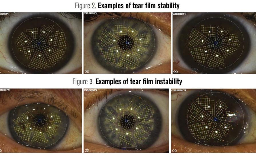
An unstable tear film introduces optical irregularity between blinks, distorting keratometry and wavefront aberrometry. This tear film instability induces artifactual astigmatism, potentially leading to misplaced steep-axis incisions or inappropriate toric lens selection. Meanwhile, fluctuating corneal curvatures introduce errors into the baseline K-values, which directly translate to errors in the final intraocular lens (IOL) power calculations.
This instability is not confined to a single sitting either; the same unsettled surface can produce different readings from blink to blink within one exam and from visit to visit days apart, undermining the reproducibility biometry depends on. Direct evidence for this comes from a 2024 study that repeated biometry in the same cataract patients days apart and found the planned IOL power changed in a quarter of cases purely because of measurement variability, with more marked dry eye signs driving the discrepancy. The same instability affects higher-order aberration measurement, with studies linking tear film quality directly to aberrometric readings. So an unsettled surface distorts not just the numbers used for standard IOL selection, but the finer optical data premium lens planning increasingly depends on. The downstream consequence is a heightened risk of refractive surprise after an otherwise technically successful operation, a risk best measured against the standard the profession sets for itself. The Royal College of Ophthalmologists considers biometry outcomes excellent only when at least 55 per cent of eyes land within ±0.50 dioptres of target postoperatively, a bar undiagnosed ocular surface disease makes considerably harder to clear, particularly for toric, multifocal, and refractive surgery candidates, where premium pricing brings premium expectations and even small errors are poorly tolerated.
The relationship also runs in the opposite direction. Surgery itself is a recognised cause of DED, not merely a setting that exposes pre-existing disease. Corneal and limbal incisions sever corneal sensory nerves, disrupting the sensory-secretory feedback loop described earlier and producing a transient neurotrophic reduction in reflexive tearing and blink rate, an effect documented after laser refractive procedures as well as cataract surgery. Reduced and altered blinking during recovery limits tear distribution, while preservatives in postoperative drops and prolonged antibiotic and steroid courses add further surface toxicity.
Because this reverse relationship feeds straight back into the measurement problem described earlier, leaving DED undiagnosed until after surgery threatens the very outcome the surgery is trying to protect. Preoperative treatment has been shown to cut postoperative refractive surprise rates from over 15 per cent to under 4 per cent, evidence that the ocular surface must be identified and optimised before the eye is ever measured for surgery.
Preoperative assessment
Because unrecognised ocular surface disease can directly undermine the measurements described above, preoperative screening should be considered a routine part of the cataract and refractive workup rather than an occasional add-on. This is compounded by the demographics of the cataract population itself: DED increases in prevalence with age, driven by declining tear production and meibomian gland dropout, meaning the average cataract candidate is already in a group where undiagnosed disease is more likely. Much of this goes undiagnosed, often masked by the visual complaints attributed to the cataract itself. A survey of ophthalmologists found that one in three were not routinely assessing patients for dry eye before cataract surgery.
A structured approach should begin with a history and review of risk factors, covering age, female sex, contact lens wear, prior refractive surgery, systemic disease such as diabetes or autoimmune disorders, and medications known to disrupt the tear film. Alongside this, a validated symptom questionnaire should be completed. The Ocular Surface Disease Index (OSDI) remains the most widely used, with the shorter OSDI-6 and the Standard Patient Evaluation of Eye Dryness (SPEED) offering quicker alternatives, and the American Society of Cataract and Refractive Surgery’s SPEED II preoperative version was designed specifically for this setting. Because symptom scores and objective signs are often poorly correlated, particularly in older patients whose corneal sensitivity has declined, a questionnaire alone is not sufficient and must be paired with clinical examination.
The clinical exam should assess lid position and closure, blink rate and completeness, and gross corneal sensation, since reduced sensitivity blunts the reflex blink and tearing response and often goes unnoticed by the patient. Tear meniscus height gives a quick estimate of aqueous volume, while gentle pressure on the lower lid margin reveals meibum quality and identifies meibomian gland dysfunction that might otherwise be missed. Tear breakup time and fluorescein or lissamine green staining should be performed in every patient, as they are quick to obtain and directly assess tear film stability and epithelial integrity, the two parameters most closely tied to keratometric and biometric error described earlier.
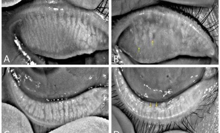
Where these initial tests raise concern, or where point-of-care devices are available, testing should extend to tear osmolarity and matrix metalloproteinase-9 (MMP-9), which mark hyperosmolarity and surface inflammation respectively, and to meibography, which images gland structure and severity of dropout beyond what lid expression alone can show.
Outcome
The outcome of this workup should guide a clear decision rather than a vague impression of surface health. If screening is negative, surgery can proceed as planned. If mild ocular surface disease is present, but not judged visually significant, surgery can also proceed, with patient counselling and prophylactic treatment to reduce the risk of postoperative symptoms. Where signs are more marked, particularly abnormal tear osmolarity or MMP-9, unstable topography or significant staining, surgery should be deferred until targeted treatment has been given time to work, with reassessment typically at two to four weeks. It is this treatment phase, and the evidence behind each intervention, that follows next.
Once assessment has identified ocular surface disease that requires treatment, the choice of intervention should match the underlying DED subtype and the severity rather than following a fixed sequence. The evidence for each option is strongest when it is tailored to what the preoperative window actually needs to achieve: Rapid, measurable improvement in tear film stability before biometry is repeated.
For evaporative disease and meibomian gland dysfunction, lid hygiene and warm compresses remain the first practical step. A randomised trial of patients undergoing cataract surgery found that 20 minutes of warm compresses followed by lid massage before surgery improved postoperative tear film stability. In practice, real-world adherence to home-based warm compress regimens is frequently inadequate when performed by patients themselves, particularly in an older surgical population. An alternative option, developed by Mr Michael Gallagher to address inconsistent heat delivery, is the Eye Spoon, a wooden heat therapy device which delivers targeted and sustained heat to the eyelid margin.

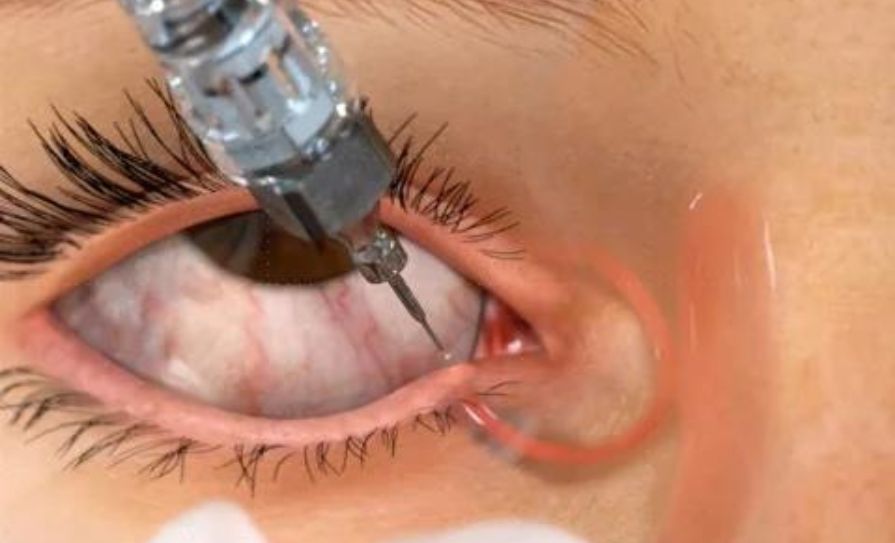
Preservative-free artificial tears remain the foundation of treatment for most patients regardless of subtype, and should be used at a frequency of three to four times daily rather than once or twice, since less frequent dosing is unlikely to be sufficient. Preservative-containing drops and topical non-steroidal anti-inflammatories should be avoided where possible in the preoperative window since benzalkonium chloride, the preservative present in most multidose ophthalmic formulations, is well documented to damage corneal and conjunctival epithelial cells and further destabilise the tear film.
Where inflammation is a significant driver, a short course of topical corticosteroid or an immunomodulatory agent such as ciclosporin or lifitegrast can be considered. Evidence suggests that the duration of treatment matters: A three-day course of corticosteroid showed no measurable benefit in one trial, whereas courses of four weeks or longer produced significant improvement in both signs and patient-reported symptoms. Ciclosporin and lifitegrast have shown similar benefit over a comparable preoperative timeframe, with objective improvement in tear film stability tracking alongside improvement in validated symptom scores, giving reasonable confidence that improving one is not achieved at the expense of the other. Corticosteroids nonetheless carry a well-recognised side-effect profile, including elevated intraocular pressure even in short courses of a few weeks and cataract progression with prolonged use. Their role here is ideally as a brief, targeted course to settle inflammation ahead of surgery rather than as ongoing therapy.
For patients with aqueous deficiency or inadequate response to artificial tear replacement, punctal occlusion can help retain what tear volume is present. Traditional punctal plugs can be effective, but recognised drawbacks include epiphora, foreign body sensation and spontaneous loss of the plug, while intracanalicular designs in particular carry a risk of migration and canaliculitis. A newer option is Lacrifill, a crosslinked hyaluronic acid canalicular gel, which fills the entire length of the canalicular lumen rather than merely sitting within the opening. Because this leaves no space within the canalicular system for tears to stagnate, it carries a lower risk of infection, and clinical trial data show comparable or superior improvement in tear volume and surface staining sustained out to six months.
For obstructive meibomian gland dysfunction unresponsive to home measures, in-office thermal pulsation therapy can restore glandular patency and lipid layer stability more reliably than compresses alone. When performed several weeks before surgery, it has been shown to improve meibomian gland secretion and tear breakup time preoperatively, alongside more accurate keratometry and IOL calculation, though its effect on subjective symptom scores is more variable than its effect on objective signs, a discrepancy worth bearing in mind when counselling patients on what to expect. Intense pulsed light combined with gland expression has shown similar promise for refractive accuracy in this population, though the evidence base remains smaller.
Conclusion
Across every subtype and severity, the preoperative goal is the same: Targeted correction of whatever specific deficit the assessment has identified, delivered on a timeline the surgical booking can accommodate. Lid hygiene, artificial tears, anti-inflammatory therapy, punctal occlusion, and thermal pulsation each address a different point in the tear film cycle, but their shared purpose is to restore stability before the eye is re-measured and surgical intervention begins.
DED occupies an uncomfortable position in cataract and refractive practice, often treated as a minor comfort issue when it is in fact a direct threat to measurement accuracy and refractive outcome. The tear film’s role as the eye’s outermost optical surface means its instability propagates through keratometry, aberrometry, and IOL power calculation, while surgery itself perpetuates the problem through nerve transection, altered blinking and drop toxicity. Breaking this cycle depends on treating preoperative screening as routine rather than incidental, given how often ocular surface disease in this population is masked by symptoms attributed to the cataract itself. Once identified, treatment should be matched to subtype and severity rather than applied as a fixed sequence, with the shared aim of restoring tear film stability within a timeframe the surgical booking can accommodate. For ophthalmologists managing an ageing surgical population, incorporating this assessment into standard preoperative pathways offers a meaningful and achievable opportunity to reduce refractive surprise and improve patient satisfaction after otherwise technically successful surgery.
References
Craig JP, et al. (2017) TFOS DEWS II definition and classification report, The Ocular Surface, 15(3), pp. 276–283. Available at: www.tfosdewsreport.org/report-definition_and_classification/48_36/en/
Manzouri B, Ahmad S, and Harper S et al. (2026) Assessment and management of dry eye disease in the UK: Standardising reality-based best practice, Eye, 40, pp. 1185–1195. Available at: https://doi.org/10.1038/s41433-026-04375-7
Basilious A, Xu CY, and Malvankar-Mehta MS. (2022) Dry eye disease and psychiatric disorders: A systematic review and meta-analysis, European Journal of Ophthalmology, 32(6), pp. 3199–3212. Available at: https://journals.sagepub.com/doi/full/10.1177/11206721211060963
Koh S. (2018) Mechanisms of visual disturbance in dry eye, Cornea, 37(Suppl 1), pp. S30–S33. Available at: www.ovid.com/jnls/corneajrnl/abstract/10.1097/ico.0000000000000998~mech anisms-of-visual-disturbance-in-dry-eye
Morthen MK, Magno MS, Utheim TP, Hammond CJ, and Vehof J. (2023) The work-related burden of dry eye, Ocular Surface, 28, pp. 30–36. Available at: https://doi.org/10.1016/j.jtos.2023.01.006
Forrester JV and Pearlman E et al. (2016) Tear film, in the eye (Fourth Edition). Amsterdam: Elsevier. Available at: www.sciencedirect.com/science/article/pii/B9780702055546000046
Nagstrup AH. (2023) The use of benzalkonium chloride in topical glaucoma treatment, acta ophthalmologica, 101(Suppl. 278), pp. 3–21. Available at: https://doi.org/10.1111/aos.15808
Al-Aqaba MA et al. (2019) Corneal nerves in health and disease, progress in retinal and eye research, 73, p. 100762. Available at: https://pmc.ncbi.nlm.nih.gov/articles/PMC5872636/
Vidal-Rohr M, Craig JP, Davies LN, and Wolffsohn JS. (2024) Classification of dry eye disease subtypes, Contact Lens and Anterior Eye, 47(5), 102257. Available at: https://doi.org/10.1016/j.clae.2024.102257
Stapleton F et al. (2017) TFOS DEWS II Epidemiology Report, The Ocular Surface, 15(3), pp. 334–365. Available at: www.tfosdewsreport.org/report-epidemiology_report/71_36/en/
Wolffsohn JS et al. (2021) The relationship between dry eye disease and digital screen use, Clinical Ophthalmology, 15, pp. 3811–3820. Available at: www.dovepress.com/the-relationship-between-dry-eye-disease-and-digital-screen-use-peer-reviewed-fulltext-article-OPTH
Coco G et al. (2025) A practical approach for optimising ocular surface status before cataract surgery to improve visual outcomes and reduce the risk of postoperative dry eye, Ophthalmology and Therapy, 14, pp. 2697–2733. Available at: https://doi.org/10.1007/s40123-025-01251-7
Matossian C. (2020) How the tear film affects IOL measurements, Optometry Times, August 2020, Volume 12, Issue 8. Available at: www.optometrytimes.com/view/how-the-tear-film-affects-iol-measurements
Ahn S, Eom Y, Song JS, and Kim DH. (2024) Short-term variability in ocular biometry and the impact of preoperative dry eye, Scientific Reports, 14, p. 26762. Available at: https://doi.org/10.1038/s41598-024-77572-7
Rhee J, Chan TC, and Chow SS et al. (2022) A systematic review on the association between tear film metrics and higher order aberrations in dry eye disease and treatment, Ophthalmology and Therapy, 11(1), pp. 35–67
Thompson V, Karpuk K, and Packer M. (2025) Preoperative ocular surface optimisation and the role of lacrimal occlusion in dry eye management before cataract and refractive surgery: A critical perspective, Expert Review of Ophthalmology. Available at: https://doi.org/10.1080/17469899.2025.2606426
Nemet A, Mimouni M, and Hecht I et al. (2020) Post laser-assisted in-situ keratomileusis dry eye disease and temporary punctal plugs, Indian Journal of Ophthalmology, 68(12), pp. 2960–2963. Available at: https://doi.org/10.4103/ijo.IJO_1664_20
Kim J, Kim MK, Ha Y, Paik HJ, and Kim DH. (2021) Improved accuracy of intraocular lens power calculation by preoperative management of dry eye disease, BMC Ophthalmology, 21, pp. 1–7. Available at: https://doi.org/10.1186/s12886-021-02129-5
Bandlitz S, Bäumer J, Conrad U, and Wolffsohn JS. (2017) Usability and reproducibility of tear meniscus values generated via swept-source optical coherence tomography and the slit lamp with a graticule method, Contact Lens and Anterior Eye, 40(6), pp. 372–376. Available at: https://doi.org/10.1016/j.clae.2017.09.010
Du Y-L, Peng X, Liu Y, Wang J-S, Ye Y-F, Xu K-K, Qu J-Y, Chen H, Xie H-T, and Zhang M-C. (2023) Ductal hyperkeratinisation and acinar renewal abnormality: New concepts on pathogenesis of meibomian gland dysfunction, Current Issues in Molecular Biology, 45(3), pp.1889–1901. Available at: https://doi.org/10.3390/cimb45030122
Goldstein MH, Silva FQ, Blender N, Tran T, and Vantipalli S. (2021) Ocular benzalkonium chloride exposure: Problems and solutions, Eye, 35(11), pp. 2985–2993. Available at: https://doi.org/10.1038/s41433-021-01668-x
Liu S-H, Saldanha IJ, Abraham AG, Rittiphairoj T, Hauswirth S, Gregory D, Ifantides C, and Li T. (2022) Topical corticosteroids for dry eye, Cochrane Database of Systematic Reviews, Issue 10, Art. No. CD015070. Available at: https://doi.org/10.1002/14651858.CD015070.pub2
Ervin A-M, Law A, and Pucker AD. (2019) Punctal occlusion for dry eye syndrome: Summary of a Cochrane systematic review, British Journal of Ophthalmology, 103(3), pp.301–306. Available at: https://doi.org/10.1136/bjophthalmol-2018-312756
Packer M, Lindstrom R, Thompson V, Parekh JG, Gupta P, Nijm, LM, and Donnenfeld E. (2024) Effectiveness and safety of a novel crosslinked hyaluronate canalicular gel occlusive device for dry eye, Journal of Cataract and Refractive Surgery, 50(10), pp. 1051–1057. Available at: https://doi.org/10.1097/j.jcrs.0000000000001505
Stodola E. (2024) Punctal occlusion: A valuable resource for dry eye patients, EyeWorld, Fall issue. Available at: www.eyeworld.org/2024/punctal-occlusion-a-valuable-resource-for-dry-eye-patients/









Leave a Reply
You must be logged in to post a comment.