







Reference: July-August 2026 | Issue 4 | Vol 19 | Page 47
While silver dressings are widely accepted in modern tissue viability practice, colloidal silver remains under-recognised in formal guidelines despite strong evidence and observation
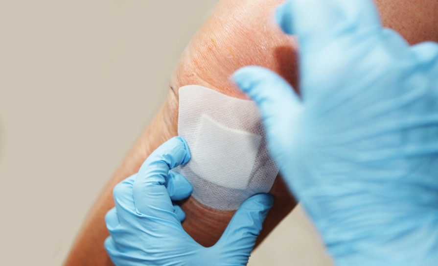
Silver’s antimicrobial use predates recorded medical history,¹ and its role in wound care remains firmly established. Today’s clinical practice commonly incorporates silver foam dressings, hydrofibres, alginates, and silver-containing creams.2,3,4 Colloidal silver, distinct from ionic forms, contains nanoscale metallic particles that exhibit antimicrobial and anti-inflammatory effects.5,6,7,8,9
Colloidal silver is typically produced at 10-100 parts per million (ppm); concentrations of 50-70ppm provide effective antimicrobial action without cytotoxicity. It is commonly used as a topical spray or gauze soak. Despite widespread community use – particularly in Germany and the Netherlands – colloidal silver remains absent from Irish clinical guidelines due to limited large-scale trials.
A narrative review of available evidence on the antimicrobial properties, wound-healing potential, safety, and clinical relevance of topical colloidal silver solutions (50-70ppm) as adjuncts to contemporary wound management was conducted using PubMed, PMC, wound-care journals, nanomedicine studies, and observational practice literature. Comparative evaluation with silver-impregnated dressings (Ag+) was included.
Types of silver used in wound care
Silver sulfadiazine (SSD): SSD has been a mainstay in burn management for decades.10 However, SSD demonstrates significant cytotoxicity to keratinocytes and fibroblasts, delaying epithelialisation.10
Silver nitrate: Historically used for infection control, silver nitrate requires frequent reapplication and can cause tissue staining.11
Silver-impregnated dressings (Ag+): Nanocrystalline silver and ionic silver dressings (eg, AquaCel Ag, Mepilex Ag) are widely studied.2,4 They provide broad antimicrobial coverage but are costly and may not fully contact the wound surface.
Colloidal silver: Colloidal silver consists of nanoscale metallic particles (10-100nm) suspended in water.6,7 Evidence suggests strong antimicrobial action with low cytotoxicity at 50-70ppm.5,6
Mechanisms of action
ANTIMICROBIAL ACTIVITY
Silver nanoparticles demonstrate activity against:
✽ Staphylococcus aureus
✽ Methicillin-resistant Staphylococcus aureus (MRSA)
✽ Pseudomonas aeruginosa
✽ Escherichia coli
✽ Anaerobes
✽ Fungi (Candida).
Mechanisms include disruption of bacterial cell membranes; interference with DNA replication; inhibition of respiratory enzymes; oxidative stress induction; and destabilisation of biofilm matrices. Multiple studies demonstrate membrane disruption, enzyme inhibition, and interference with DNA function.3,5,6,8,12 Importantly, silver nanoparticles exert multi-target antimicrobial effects, reducing the likelihood of microbial resistance development compared with single-target antibiotic therapies.8,12,13
BIOFILM DISRUPTION
Biofilms contribute significantly to wound chronicity. Silver nanoparticles penetrate and destabilise biofilm matrices, increasing bacterial susceptibility to clearance.3,4,14
ANTI-INFLAMMATORY EFFECTS
Topical nanosilver reduces inflammatory cytokines and decreases redness, odour, and exudate.6,7,15
PRESERVATION OF VIABLE TISSUE
Unlike SSD, nanoparticle silver at 50-70ppm does not damage fibroblasts.10 This preserves granulation potential and supports epithelialisation.
Mechanistic insights from German scientific literature
German scientific author Dr Josef Pies has published detailed technical reviews on the chemistry, safety, and antimicrobial mechanisms of colloidal and nanosilver. His analyses emphasise the influence of particle size, surface charge, and ion release on antimicrobial efficacy, and highlight the important distinction between true nanoparticle suspensions and purely ionic silver solutions.16
Although Pies is not a primary clinical researcher, his mechanistic summaries are grounded in peer-reviewed nanomedicine studies and support established laboratory findings demonstrating nanoparticle silver’s ability to disrupt microbial membranes, inhibit enzymatic processes, and destabilise biofilms. His work provides additional mechanistic context that aligns with contemporary experimental evidence and supports the rationale for topical colloidal silver use in wound care.16
Evidence review
In-vitro evidence: Multiple laboratory studies demonstrate that:
✽ 25-50ppm colloidal silver inhibits common wound pathogens5,6
✽ MRSA and pseudomonas are highly sensitive5,6
✽ Fungal inhibition has been demonstrated in burn pathogens2
✽ Biofilm formation is inhibited5,6
✽ Nanoparticles have a prolonged antimicrobial effect due to slow ion release.5,6 These results are consistent across nanomedicine, antimicrobial, and wound microbiology studies.5,6,8,9,12
Animal studies: Rodent models show faster epithelialisation, lower bacterial loads, and reduced inflammation compared with SSD.4
Human and clinical evidence: While large randomised trials are limited, available data report:
✽ Faster healing in chronic venous ulcers treated with colloidal silver compresses⁷
✽ Emerging evidence and clinical observations suggest potential benefit in diabetic foot and chronic venous ulcers⁷
✽ Reduced odour and exudate in heavily colonised wounds⁷
✽ Effective decontamination post-debridement⁷
✽ Enhanced comfort in patients with dermatitis-associated wounds and partial-thickness burns.⁷
Community and clinical practice observations indicate that across Europe, colloidal silver is used as a spray or soaked-gauze dressing. Noted benefits include rapid cooling, reduced bioburden, and improved granulation. It is considered particularly useful in home-care patients where dressings are limited. These observations align with laboratory evidence and provide real-world context.
Safety and contraindications
Topical safety: Short-term external use shows minimal systemic absorption.¹⁷ No cases of argyria from external application have been reported. There is a low irritation risk at 50-70ppm.
When to avoid:
✽ Deep cavity wounds (solution pooling risk)
✽ Closed abscesses
✽ Combination with iodine, hydrogen peroxide, vinegar, or acids
✽ Continuous use beyond three months without reassessment.
| FEATURE | SSD | COLLOIDAL SILVER |
|---|---|---|
| Cytotoxicity | High | None at ≤70ppm |
| Biofilm activity |
Weak | Strong |
| Application | Cream | Spray/soak |
| Cost | Moderate | Low |
| Community suitability |
Moderate | Excellent |
TABLE 1: Comparison between colloidal silver and SSD
Clinical application guidance
RECOMMENDED CONCENTRATION
A concentration of 50-70ppm is supported by mechanistic and cytotoxicity data.5,6 Below 20ppm is insufficient, while above 100ppm offers no proven benefit and increases irritation.
INDICATIONS
Indications for the use of colloidal silver include:
✽ Colonised or infected wounds
✽ Chronic venous ulcers⁷
✽ Diabetic ulcers
✽ Partial-thickness burns2
✽ Fungal or mixed bioburden wounds.2
APPLICATION METHODS
Spray method:
1. Clean wound
2. Spray liberally
3. Apply secondary dressing, ie, foam/alginate.
Gauze-soak method:
✽ Saturate sterile gauze in colloidal silver
✽ Apply directly to wound bed
✽ Cover with foam/alginate
✽ Change daily
✽ Most effective for full wound-bed contact.
DISCONTINUATION
✽ When healthy granulation is present
✽ When infection is controlled.
Limitations and concerns
Despite several limitations and ongoing controversies surrounding colloidal silver, interest in its potential therapeutic applications continues to grow. Key concerns include the lack of standardisation across commercially available products, limited large-scale randomised controlled trials, and significant variability in nanoparticle size, purity, and concentration between manufacturers – all of which can affect safety and efficacy. Regulatory authorities have also expressed caution due to the historical misuse of silver products.
Nevertheless, these limitations do not invalidate the expanding body of credible laboratory research and encouraging clinical observations demonstrating antimicrobial, wound-healing, and anti-inflammatory properties in certain formulations. As research methods improve and product quality becomes more consistent, colloidal silver remains an area of ongoing scientific interest and investigation.
Discussion
Colloidal silver demonstrates antimicrobial efficacy comparable to silver dressings5,7 and superior biofilm action.3 Its low cost and broad accessibility make it particularly suitable for community wound care settings, where dressing choice may be limited. Mechanistic reviews, including those by Pies,16 reinforce the scientific rationale for its topical use.
Colloidal silver occupies a unique position in modern wound care; it is scientifically promising, clinically useful, but under-represented in guidelines. Recent nanomedicine and wound-care literature continues to support the antimicrobial efficacy, biofilm disruption, and clinical relevance of silver nanoparticles in wound management.8,9,12,14,15,18,19
The evidence supports that:
✽ Its antimicrobial activity is comparable to silver dressings
✽ Its biofilm disruption is particularly valuable
✽ It poses minimal toxicity risk when used correctly
✽ Its low cost and ease of use make it highly suitable for community nursing and GP settings.
Observational data from experienced practitioners, including those trained in German wound-care tradition, mirror laboratory findings and offer practical insights where trials are lacking. Given the rising burden of chronic wounds, polypharmacy, community-based care, and infection control challenges, it is reasonable to consider colloidal silver a viable adjunctive wound care tool.
Conclusion
Colloidal silver at 50-70ppm is an effective, safe, and accessible adjunct for modern wound care. While more controlled trials are needed, existing evidence supports its use for infected and chronic wounds, particularly in community practice.
Integration into Irish frameworks is justified given the strength of antimicrobial data, low cytotoxicity, and positive real-world patient outcomes.
References
- Lansdown AB. Silver in health care: Antimicrobial effects and safety in use. Curr Probl Dermatol. 2006;33:17-34. doi:10.1159/000093928
- Wright JB, Lam K, Hansen D, Burrell RE. Efficacy of topical silver against fungal burn wound pathogens. Am J Infect Control. 1999;27(4):344-350. doi:10.1016/s0196-6553(99)70055-6.
- Percival SL, Bowler PG, Russell D. Bacterial resistance to silver in wound care. J Hosp Infect. 2005;60(1):1-7. doi:10.1016/j.jhin.2004.11.014.
- Fong J, Wood F. Nanocrystalline silver dressings in wound management: A review. Int J Nanomedicine. 2006;1(4):441-449. doi:10.2147/nano.2006.1.4.441.
- Rai M, Yadav A, Gade A. Silver nanoparticles as a new generation of antimicrobials. Biotechnol Adv. 2009;27(1):76-83. doi:10.1016/j.biotechadv.2008.09.002.
- Kim JS, Kuk E, Yu KN, et al. Antimicrobial effects of silver nanoparticles. Nanomedicine. 2007;3(1):95-101. doi:10.1016/j.nano.2006.12.001.
- Du Y, Lu J, Guo X, et al. The role of silver and silver-based products in wound management: A review of advances and current landscape. J Funct Biomater. 2026;17(1):27. doi:10.3390/jfb17010027.
- Qing Y, Cheng L, Li R, et al. Potential antibacterial mechanism of silver nanoparticles and the optimisation of orthopaedic implants by advanced modification technologies. Int J Nanomedicine. 2018;13:3311-3327. doi:10.2147/IJN.S165125.
- Xu L, Wang YY, Huang J, et al. Silver nanoparticles: Synthesis, medical applications, and biosafety. Theranostics. 2020;10(20):8996-9031. doi:10.7150/thno.45413.
- Klasen HJ. A historical review of the use of silver in the treatment of burns. II. Renewed interest for silver. Burns. 2000;26(2):131-138. doi:10.1016/s0305-4179(99)00116-3.
- Russell AD, Hugo WB. Antimicrobial activity and action of silver. Prog Med Chem. 1994;31:351-370. doi:10.1016/s0079-6468(08)70024-9.
- Durán N, Durán M, de Jesus MB, et al. Silver nanoparticles: A new view on mechanistic aspects on antimicrobial activity. Nanomedicine. 2016;12(3):789-799. doi:10.1016/j.nano.2015.11.016.
- Singh P, Pandit S, Mokkapati V, et al. Silver nanoparticles: Biological synthesis and antimicrobial applications. J Nanobiotechnol. 2021;19:84. doi:10.1186/s12951-021-00807-1.
- Paladini F, Pollini M. Antimicrobial silver nanoparticles for wound healing application: Progress and future trends. Materials (Basel). 2019;12(16):2540. doi:10.3390/ma12162540.
- Burdușel AC, Gherasim O, Grumezescu AM, et al. Biomedical applications of silver nanoparticles: An up-to-date overview. Nanomaterials (Basel). 2018;8(9):681. doi:10.3390/nano8090681.
- Pies J. Colloidal silver: Properties, mechanisms, and applications. 4th ed. Kopp Verlag; 2018.
- Wijnhoven SWP, Peijnenburg WJGM, Herberts CA, et al. Nano-silver – a review of available data and knowledge gaps in human and environmental risk assessment. Nanotoxicology. 2009;3(2):109-138. doi:10.1080/17435390902725914.
- Leaper D. Appropriate use of silver dressings in wounds: International consensus document. Int Wound J. 2012;9(5):461-464. doi:10.1111/j.1742-481X.2012.01091.x.
- Swanson T, Ousey K, Haesler E, et al. IWII wound infection in clinical practice consensus document: 2022 update. J Wound Care. 2022;31(Sup12):S10-S21. doi:10.12968/jowc.2022.31.Sup12.S10.







Leave a Reply
You must be logged in to post a comment.