NOTE: By submitting this form and registering with us, you are providing us with permission to store your personal data and the record of your registration. In addition, registration with the Medical Independent includes granting consent for the delivery of that additional professional content and targeted ads, and the cookies required to deliver same. View our Privacy Policy and Cookie Notice for further details.
LEARNING OVERVIEW This educational module explores the critical role of imaging in lung cancer screening, with a particular focus on the use of low dose computed tomography (LDCT) in high risk populations. The module highlights the global burden of lung cancer, the rationale for early detection, and the evidence supporting structured screening programmes. It also examines the benefits and limitations of screening, outlines current eligibility criteria, and discusses emerging developments, such as artificial intelligence (AI) and advanced imaging technologies.
This module is primarily intended for radiology healthcare professionals, including radiographers and radiology residents. It is also relevant to radiologists and other clinicians involved in lung cancer screening pathways. Upon completion, learners should be able to understand the evidence base for LDCT screening, apply appropriate screening criteria, recognise the limitations of imaging, and appreciate their role in improving patient outcomes.
Introduction
Lung cancer continues to represent a major global health challenge. According to the World Health Organization (WHO), lung cancer accounts for approximately 1.8 million deaths annually, making it the leading cause of cancer-related mortality worldwide.1 Despite advancements in oncological therapies, including targeted treatments and immunotherapy, overall survival rates remain relatively poor. This reflects the fact that lung cancer is frequently diagnosed at an advanced stage, at which point curative treatment options are limited.
The asymptomatic nature of early-stage lung cancer presents a significant barrier to timely diagnosis. Patients typically develop symptoms such as persistent cough, haemoptysis, or weight loss only when the disease has progressed. Consequently, there has been increasing emphasis on the development and implementation of screening strategies aimed at detecting lung cancer in asymptomatic individuals who are at high risk. Imaging plays a central role in these screening programmes, providing a non-invasive method for identifying early pathological changes within the lungs. The introduction of LDCT has been particularly transformative, enabling the detection of small pulmonary nodules that would otherwise remain undetected.
Over the past two decades, a growing body of evidence has demonstrated that LDCT screening can significantly reduce lung cancer mortality in high risk populations. This has led to the adoption of national screening programmes in several countries and has reinforced the importance of imaging in preventive health care. This module examines the role of imaging in lung cancer screening, with a focus on the rationale for screening, the evolution of screening methods, the evidence base supporting current practices, and the critical role of radiology professionals in delivering effective screening services.
Rationale for screening
The rationale for lung cancer screening is grounded in the well established relationship between stage at diagnosis and patient survival. Lung cancer is typically classified into stages based on tumour size, lymph node involvement, and the presence of metastases. Survival outcomes vary dramatically across these stages, with early stage associated with significantly better prognosis.
Patients diagnosed with stage I lung cancer have a five-year survival rate that may exceed approximately 60 to 70 per cent, depending on cohort and treatment modality, particularly when treated with surgical resection or stereotactic radiotherapy.2 In contrast, patients with stage IV, characterised by distant metastases, have a five-year survival rate of less than 10 per cent.2 This stark contrast underscores the critical importance of detecting lung cancer at an early, potentially curable stage.
Screening aims to identify disease before the onset of symptoms, thereby shifting the stage distribution toward earlier stages. This is, often referred to as stage migration and has a direct impact on overall survival rates within the screened population. Early detection also facilitates the use of less invasive treatment, reduces treatment-related morbidity, and improves quality of life. In addition to clinical benefits, early detection has important economic implications.
The cost of treating advanced lung cancer, which often involves complex multimodal therapies, is substantially higher than that of treating early stage. Consequently, effective screening programmes have the potential to reduce healthcare expenditure in the long term, although cost effectiveness depends on population selection, adherence, and healthcare system factors, provided that they are appropriately targeted and efficiently implemented.3
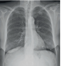
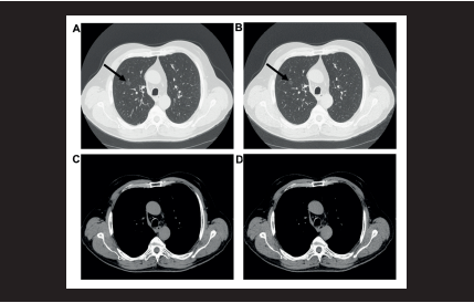
FIGURE 1: Lung cancer – missed on initial chest x-ray. Case courtesy of Philipp Widmayer, Radiopaedia.org, rID: 151887FIGURE 2: 76-year-old male (body weight 73kg) who underwent chest CT as a follow-up study for a ground-glass pulmonary nodule. (a and c; standard dose images. b and d; low dose images). A purely ground-glass nodule in right upper lobe was clearly visualised both with standard dose CT and low dose CT (arrow). Source: https://doi.org/10.1016/j.ejro.2016.04.001
The identification of high risk populations is a key component of screening strategies. Tobacco smoking and age remains the most significant risk factor for lung cancer, accounting for the majority of cases. As such, screening programmes are typically directed at current or former smokers with a significant smoking history.
By focusing on individuals with the highest risk, screening programmes aim to maximise the benefit to harm ratio. However, risk prediction models such as the 2012 Prostate, Lung, Colorectal, and Ovarian Cancer screening trial model (PLCOm2012) have been shown to identify higher risk individuals who may not meet standard eligibility criteria, detecting more lung cancers than age and smoking history criteria alone.4
Screening methods
Chest x-ray: Historical context and limitations Chest radiography (CXR) was historically considered a potential tool for lung cancer screening due to its widespread availability, low cost, and relatively low radiation dose. However, large scale studies conducted over several decades failed to demonstrate a meaningful reduction in lung cancer mortality associated with chest x-ray screening.4
The limitations of CXR is primarily related to its relatively low sensitivity for detecting small pulmonary nodules. The two-dimensional nature of the image results in the superimposition of anatomical structures, which can obscure lesions, particularly those located in complex regions such as the mediastinum or lung apices. Furthermore, CXR are less effective at identifying early stage cancers, which are often small and subtle in appearance (Figure 1).
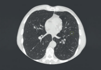
LDCT The introduction of LDCT marked a significant advancement in lung cancer screening. Unlike CXR, CT imaging provides cross sectional images of the thorax, allowing for detailed visualisation of lung parenchyma without the issue of anatomical overlap. LDCT protocols are specifically designed to minimise radiation exposure while maintaining sufficient image quality for the detection of small nodules (Figures 2, 3).
FIGURE 3: Axial CT image in a 63-year-old male participant shows a typical perifissural nodule with triangular shape. This perifissural nodule was detected at baseline, with a volume of 61mm3. Source: https://doi.org/10.1148/radiol.240535
Typically, LDCT scans deliver a radiation dose in the range of approximately 1 to 2 millisieverts, depending on protocol and patient factors, which is substantially lower than that of standard diagnostic CT scans.5 This reduction is achieved through the use of lower tube current settings, reduced voltage, and advanced image reconstruction techniques. These technological advancements have enabled the widespread adoption of LDCT in screening programmes.5
LDCT is highly sensitive for the detection of pulmonary nodules, including those measuring less than 10 millimetres in diameter. In addition to detecting nodules, CT imaging allows for the assessment of nodule characteristics, such as size, shape, and density, which are important for risk stratification. Volumetric analysis and growth rate assessment are also possible, facilitating the differentiation between benign and malignant lesions.6,7,8
The implementation of LDCT screening requires standardised protocols to ensure consistency and reliability. Reporting systems such as the Lung Imaging Reporting and Data System, developed by the American College of Radiology (ACR), provide a structured classification system. Categories range from 1 (negative, no nodules or definitely benign findings) through to 4 (suspicious, further investigation warranted), with management recommendations linked to each category to guide clinical decision making (Table 1). This standardisation helps reduce inter-observer variability and improves the efficiency and safety of screening programmes.9
Evidence supporting screening
The adoption of LDCT as the primary modality for lung cancer screening is supported by robust evidence from large randomised controlled trials. Among these, the National Lung Screening Trial (NLST) and the NELSON trial are particularly influential. The NLST was a landmark study conducted in the United States, involving more than 53,000 participants who were at high risk for lung cancer. Participants were randomly assigned to undergo annual screening with either LDCT or CXR. The results demonstrated approximately a 20 per cent reduction in lung cancer mortality in major trials in the LDCT group compared to the CXR group.6 This finding provided the first definitive evidence that imaging based screening could reduce lung cancer mortality.
Classification criteria
Description
Atypical pulmonary cysts
New classification and management recommendations for thick-walled, multilocular cysts, and cysts with associated nodules
Juxtapleural nodules
Updated classification and management recommendations for juxtapleural nodules (perissural, costal pleural, perimediastinal, and peridiaphragmatic)
Inflammatory or infectious findings
Updated classification and management recommendations for findings that may represent an infectious or inflammatory process: Segmental or lobar consolidation, multiple new nodules (more than six in number), large solid nodules (8mm) appearing in a short interval, or new nodules in certain clinical contexts (eg, immunocompromised patient)
Airway nodules
Updated classification and management recommendations for airway nodules based on location, morphology, number, and persistence
Clarifications
Description
Growth
Updated definition: An increase >1.5mm in mean diameter within a 12-month interval
Slow growing
New definitions for slow-growing solid, part-solid, and ground glass nodules with associated management recommendations
S modifier
New guidance for when to use and discontinue use of the S modifier for potentially significant or significant findings
Management considerations
Description
Stepped management
New stepped management approach for Lung-RADS categories 3 and 4A nodules that are stable or decreasing in size at follow-up
Interval diagnostic CTs
New guidance on managing interval diagnostic CTs obtained in LCS patients, including when a diagnostic CT may substitute for an LCS examination and whether the timing of subsequent LCS imaging should be modified based on an interval diagnostic chest CT
LDCT = low-dose CT; Lung-RADS = Lung CT Screening Reporting and Data System
TABLE 1: Summary of updates in Lung-RADS v2022 Source: ACR Lung-RADSv2022: Assessment categories and management recommendations; 10.1016/j.jacr.2023.09.009
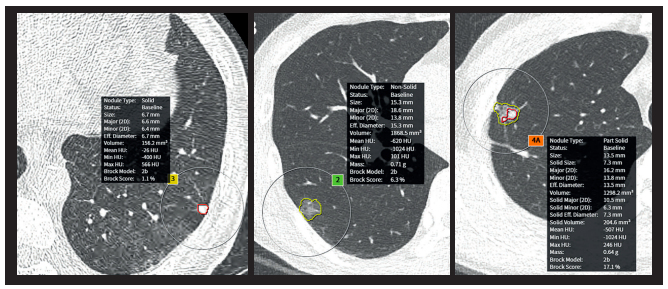
FIGURE 4: Examples of automated segmentation and measurement of lung nodules at screening LDCT Source: AI applications for thoracic imaging: Considerations for best practice
The NELSON trial, conducted in Europe, further strengthened the evidence base. This trial utilised a volumetric approach to nodule assessment and included a longer follow-up period. The results showed an approximately 24 per cent reduction in men and up to 33 per cent in women, depending on the follow-up period and analysis.7 The NELSON trial also demonstrated improved specificity compared to earlier studies, highlighting the importance of refined protocols and risk stratification methods.
These trials collectively established LDCT as an effective screening tool and provided the foundation for current clinical guidelines. Importantly, they also highlighted the need for structured follow-up protocols and multidisciplinary management to ensure optimal outcomes.
Screening criteria
The effectiveness of lung cancer screening is highly dependent on appropriate patient selection. Screening individuals at low risk may result in unnecessary investigations and potential harm, whereas targeting high risk populations maximises the likelihood of benefit. Current guidelines generally recommend screening for individuals within a specific age range who have a significant history of tobacco use.
For example, the US Preventive Services Taskforce recommends annual LDCT screening for adults aged 50 to 80 years who have a smoking history of at least 20 pack-years and who either currently smoke or have quit within the past 15 years.10 In the UK, the NHS Lung Cancer Screening Programme similarly targets high risk individuals, typically those aged 55 to 74 years who are current or former smokers, with eligibility further refined through risk assessment models to identify those most likely to benefit from LDCT screening.11
In addition to age and smoking history, other factors may influence risk, including occupational exposures, environmental factors, and underlying lung disease. There is growing interest in the use of risk prediction models that incorporate multiple variables to provide a more personalised assessment of risk. These models have the potential to improve the efficiency of screening programmes by identifying individuals who are most likely to benefit.8 The implementation of screening criteria must also consider patient comorbidities and life expectancy. Screening is generally not recommended for individuals who are unlikely to tolerate curative treatment or who have a limited life expectancy due to other conditions.
Benefits and limitations
Lung cancer screening with LDCT offers significant benefits, particularly in terms of early detection and mortality reduction. By identifying cancers at an earlier stage, screening increases the likelihood of curative treatment and improves overall survival rates. The ability to detect small nodules also enables closer monitoring and timely intervention when necessary.
However, lung cancer screening is not without limitations. One challenge is the occurrence of false positive findings, where scans detect nodules that are ultimately benign. While most positive findings do not represent cancer, structured screening protocols such as Lung-RADS help minimise unnecessary follow-up.
Nevertheless, false positives can still lead to additional imaging, occasional invasive procedures, and patient anxiety. It is important to interpret false positive statistics carefully. A positive screening result that later proves not to be cancer is not the same as the likelihood of an individual screening scan generating a false positive result. Distinguishing between these measures is important for accurately communicating screening outcomes.12
Overdiagnosis is another important concern, with estimates varying widely across studies depending on methodology and follow-up duration. Some cancers detected through screening may be slow growing and unlikely to cause harm during the patient’s lifetime. The identification and treatment of such lesions can result in unnecessary interventions and associated morbidity.
Radiation exposure is also a consideration, particularly given the repeated nature of annual screening. Although the dose associated with LDCT is relatively low, cumulative exposure over time may increase the risk of radiation-induced malignancy. Therefore, dose optimisation and adherence to appropriate screening intervals are essential, with cost and resource allocation representing additional challenges. Screening programmes require significant investment in infrastructure, equipment, healthcare professionals and staff. Ensuring equitable access to screening services is also an important consideration, particularly in resource limited settings.
Future directions
The field of lung cancer screening is evolving rapidly, with several emerging developments expected to enhance its effectiveness. One of the most promising areas is the integration of artificial intelligence (AI) into imaging workflows. AI algorithms have demonstrated the ability to assist in nodule detection, with evidence showing improvements in diagnostic performance in controlled settings. These tools have the potential to reduce workload, improve diagnostic accuracy, and decrease variability between readers (Figure 4).
Another important development is the refinement of risk-prediction models. By incorporating a wider range of variables, including genetic and environmental factors, these models aim to provide a more personalised approach to screening. This could improve the efficiency of screening programmes and reduce unnecessary investigations. Advances in imaging technology, including reductions in radiation dose, are also likely to play a significant role, particularly given the repeated nature of annual screening.
Conclusion
Imaging is central to the success of lung cancer screening programmes, with LDCT established as the modality of choice. The evidence from large-scale trials demonstrates that LDCT screening can significantly reduce lung cancer mortality in high risk populations. While screening offers substantial benefits, it also presents challenges that must be carefully managed. Radiographers play a vital role in ensuring the quality, safety, and effectiveness of screening programmes. As technology continues to advance, the integration of AI and personalised risk assessment is expected to further enhance the impact of lung cancer screening. The continued development and refinement of imaging based screening strategies will be essential in reducing the global burden of lung cancer.
References
World Health Organization. Cancer fact sheets: Lung cancer. Geneva: WHO; 2026. Available at: https://www.who.int/news-room/fact-sheets/detail/lung-cancer.
Siegel RL, Giaquinto AN, Jemal A. Cancer statistics, 2024. CA Cancer J Clin. 2024;74(1):17-48.
Black WC, Gareen IF, Soneji SS, Sicks JD, Keeler EB, Aberle DR, et al. Cost-effectiveness of CT screening in the National Lung Screening Trial. N Engl J Med. 2014;371(19):1793-1802.
Tammemägi MC, Katki HA, Hocking WG, et al. Selection criteria for lung-cancer screening. N Engl J Med. 2013;368(8):728-736.
American College of Radiology. ACR–STR practice parameter for the performance and reporting of lung cancer screening CT. Reston (VA): American College of Radiology; 2022.
Aberle DR, Adams AM, Berg CD, Black WC, Clapp JD, Fagerstrom RM, et al. Reduced lung cancer mortality with low dose computed tomographic screening. N Engl J Med. 2011;365(5):395-409.
de Koning HJ, van der Aalst CM, de Jong PA, Scholten ET, Nackaerts K, Heuvelmans MA, et al. Reduced lung-cancer mortality with volume CT screening in a randomised trial. N Engl J Med. 2020;382(6):503-513.
MacMahon H, Naidich DP, Goo JM, Lee KS, Leung ANC, Mayo JR, et al. Guidelines for management of incidental pulmonary nodules detected on CT images. Radiology. 2017;284(1):228-243.
American College of Radiology. Lung CT Screening Reporting and Data System (Lung-RADS®) Version 2022. Reston (VA): ACR; 2022. Available at: https://www.acr.org/Clinical-Resources/Clinical-Tools-and-Reference/Reporting-and-Data-Systems/Lung-RADS.
US Preventive Services Taskforce, Krist AH, Davidson KW, Mangione CM, Barry MJ, Cabana M, et al. Screening for lung cancer: US Preventive Services Taskforce recommendation statement. JAMA. 2021;325(10):962-970.
National Health Service. Lung cancer screening. Leeds: NHS England; 2023 Available at: https://www.nhs.uk/conditions/lung-cancer-screening/.
Tupper HI, Shrager JB, Moghanaki D, Simone CB 2nd, Kazerooni EA, Hart EM, et al. Misinformation and overestimation of CT lung cancer screening harms: methodology matters. J Am Coll Radiol. 2026;S1546-1440(25)00741-0.
Start this Module to earn CPD Points today
Click the "Start Module" button below
Module Title
The role of imaging in lung cancer screening programmes