



Reference: August 2026 | Issue 8 | Vol 12 | Page 53
Hepatopulmonary syndrome (HPS) is an important pulmonary vascular complication of chronic liver disease and portal hypertension. Its presentation is variable, and resting oxygen saturation alone may underestimate clinically relevant gas-exchange impairment. This review outlines a practical approach to recognising, investigating and managing HPS, with particular attention to the six-minute walk test (6MWT).
The 6MWT should not be regarded as a stand alone diagnostic test, but it can reveal exertional desaturation and prompt definitive assessment with arterial blood gas (ABG) analysis and contrast echocardiography. Early recognition matters because supportive medical therapy is limited, whereas liver transplantation can reverse the syndrome in most appropriately selected patients.
Introduction
Breathlessness in cirrhosis has a long differential diagnosis. Anaemia, ascites, hepatic hydrothorax, infection, pulmonary embolism, cardiac disease, and simple deconditioning are encountered far more often than HPS. That familiarity can work against the diagnosis: HPS may be overlooked precisely because there are several easier explanations for dyspnoea in advanced liver disease.
HPS is defined by three components: Liver disease or portal hypertension, abnormal arterial oxygenation, and evidence of intrapulmonary vascular dilatation. The International Liver Transplant Society guideline uses an elevated alveolar-arterial oxygen gradient (A-a gradient) of at least 15mmHg, or more than 20mmHg in older patients, in the appropriate clinical setting.1
HPS can occur with cirrhosis of many aetiologies and, less commonly, with portal hypertension in the absence of established cirrhosis. Its presence is not reliably predicted by the conventional severity of liver biochemical abnormalities.
Reported prevalence varies because studies have used different oxygenation thresholds and different populations. Specialist series of cirrhosis and transplant candidates commonly report HPS in the region of 10-30 per cent, while the prevalence of isolated intrapulmonary vascular dilatation is higher.2,3 This distinction matters: A positive bubble study alone is not synonymous with clinically significant HPS. Abnormal oxygenation must also be demonstrated.
It is equally important to separate HPS from portopulmonary hypertension (PoPH). Both occur in the setting of liver disease or portal hypertension, but the pulmonary physiology is very different. HPS is dominated by pulmonary vascular dilatation and shunting. PoPH is a form of pulmonary arterial hypertension characterised by increased pulmonary vascular resistance.1 The distinction changes the investigation, treatment and transplant conversation.
Why does hypoxaemia occur?
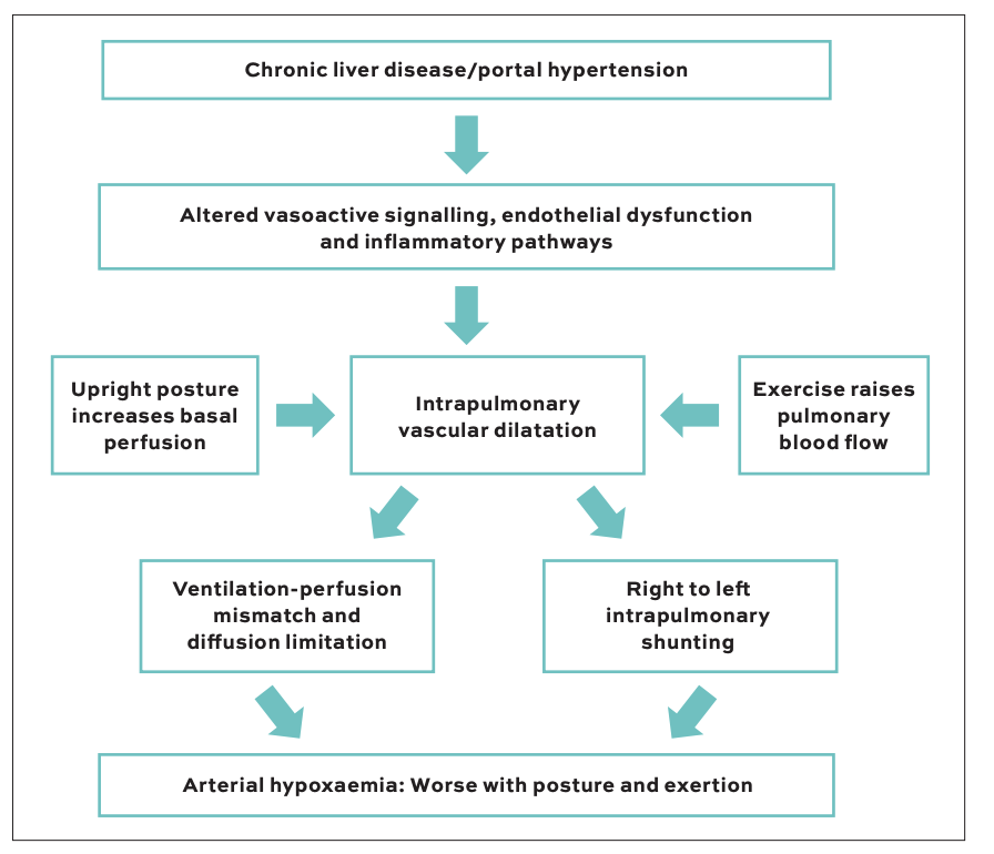
The central lesion in HPS is abnormal dilatation of the pulmonary microcirculation. Chronic liver disease alters vasoactive, endothelial, and inflammatory signalling. Increased nitric oxide-related vasodilatation, recruitment of pulmonary monocytes and macrophages, and abnormal angiogenic pathways are among the mechanisms implicated.2 The resulting capillaries may become sufficiently enlarged that oxygen cannot diffuse effectively from alveolus to haemoglobin before blood leaves the pulmonary vascular bed.
Gas exchange is therefore impaired through a mixture of ventilation-perfusion mismatch, diffusion limitation, and right to left intrapulmonary shunting. A hyperdynamic circulation can worsen the problem by shortening capillary transit time. The abnormalities tend to be more prominent at the lung bases; when the patient stands, gravitational redistribution increases blood flow to these regions. This provides a physiological explanation for platypnoea, or breathlessness that worsens upright, and orthodeoxia, a fall in oxygenation on standing. Exercise can expose the same limited oxygen reserve by increasing pulmonary blood flow and tissue oxygen demand.1,2
Presentation
The clinical spectrum is broad. Some patients have only mild gas-exchange abnormalities, whereas others develop disabling exertional dyspnoea, cyanosis, and severe resting hypoxaemia. Digital clubbing, spider naevi, platypnoea, and orthodeoxia may provide useful clues, but their absence does not exclude HPS. Symptoms are often non-specific and may be attributed to anaemia, sarcopenia, ascites, or frailty.
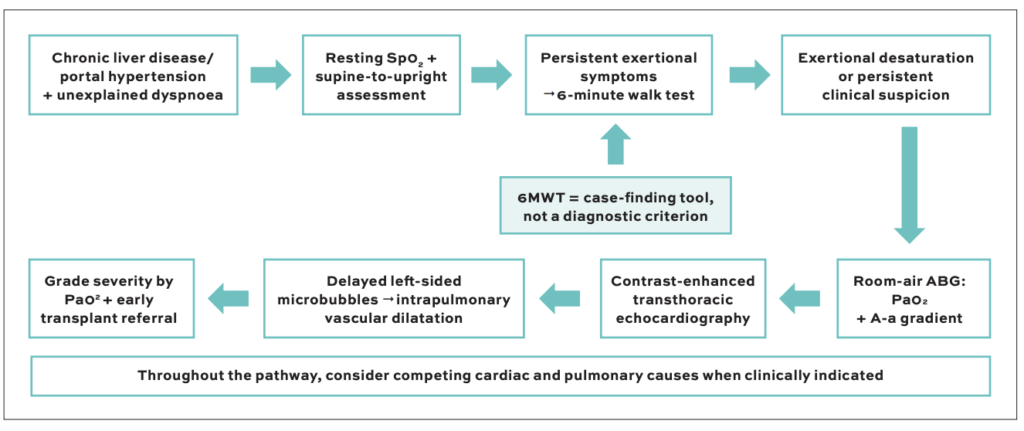
The practical difficulty is that symptoms may begin with exercise. A patient who is comfortable in bed or sitting in clinic may have a resting SpO2 in the mid-90s and still desaturate substantially when upright or walking. Pulse oximetry is therefore useful as an initial screen. Comparing oxygen saturation supine and upright is simple, and a marked postural fall should heighten suspicion. Resting pulse oximetry can also be affected by perfusion, skin temperature, motion, and device limitations. ABG analysis remains necessary when HPS is being formally assessed.
Practical clues that should trigger consideration of HPS include exertional dyspnoea that appears disproportionate to resting findings, the presence of platypnoea or orthodeoxia, unexpected oxygen desaturation during mobilisation or a six minute walk test, and otherwise unexplained hypoxaemia in a patient with chronic liver disease.
There is no need to perform an extensive HPS work-up in every patient with stable chronic liver disease. The threshold should be lower, however, when there is unexplained dyspnoea, reduced exercise tolerance, cyanosis, clubbing, platypnoea, orthodeoxia, or unexpectedly low oxygen saturation. Patients being evaluated for liver transplantation also warrant deliberate assessment because the diagnosis has implications for prognosis, allocation policy in some systems, and perioperative planning.1,2
A useful bedside sequence is to ask whether symptoms are positional, document resting saturation, repeat the measurement upright and then consider exertional assessment if the patient remains symptomatic despite reassuring resting values. This approach is deliberately pragmatic. It is intended to identify the patient in whom an ABG and contrast echocardiogram are justified, rather than to replace either test.
6MWT
The 6MWT is familiar as a measure of submaximal exercise capacity, but in HPS its most useful function may be simpler: It can expose a gas exchange abnormality that is not obvious at rest. Baseline SpO2 and heart rate are recorded, the patient walks for six minutes according to a standard local protocol, and the nadir SpO2, symptoms, and distance are documented. Continuous oximetry, when available, is helpful because a brief nadir can otherwise be missed.
In a cross-sectional study of 100 patients with cirrhosis, Singhai and colleagues identified 21 patients with HPS. Using the investigators’ predefined criteria, an abnormal 6MWT had a sensitivity of 95.2 per cent, specificity of 92.4 per cent, and negative predictive value of 98.7 per cent for HPS.4 These results are encouraging, but the study was small, single-centre, and included patients with advanced liver disease. They should not be interpreted as establishing the 6MWT as a diagnostic criterion or a universal screening test.
The safer clinical message is that the 6MWT is a case-finding tool. Significant or disproportionate exertional desaturation should prompt formal assessment of oxygenation and investigation for intrapulmonary vascular dilatation. A recently published Irish case report illustrated this role well: Exertional desaturation on the 6MWT prompted ABG measurement and bubble contrast echocardiography, which identified the intrapulmonary shunt.5 The walk test did not make the diagnosis; it revealed the clue that led to it.
This distinction matters because exercise limitation in cirrhosis is common and non-specific. Anaemia, sarcopenia, ascites, cardiopulmonary disease, and deconditioning can all reduce walking distance. A low distance alone does not point to HPS. The more useful signal is an otherwise unexplained fall in oxygen saturation, especially when it accompanies a compatible history or postural desaturation. Conversely, a normal 6MWT should not override strong clinical suspicion where symptoms, ABG findings, or posture testing suggest impaired gas exchange.
Diagnosis
ABG analysis remains central because it provides both PaO2 and the A-a gradient. The A-a gradient can become abnormal before the PaO2 is strikingly low and is therefore particularly useful when resting pulse oximetry is only mildly reduced. HPS severity is conventionally graded by room-air PaO2: mild at 80mmHg or above, moderate at 60-79mmHg, severe at 50-59mmHg, and very severe below 50mmHg.1 Severity grading is clinically useful because it informs prognosis, transplant prioritisation in some systems and perioperative planning.
The second requirement is demonstration of intrapulmonary vascular dilatation. Contrast-enhanced transthoracic echocardiography with agitated saline is the usual first-line test. Microbubbles normally opacify the right-sided chambers and are filtered by the pulmonary circulation. Delayed appearance in the left heart after passage through the lungs supports an intrapulmonary shunt.
Very early left-sided opacification is more suggestive of an intracardiac communication.1,2 Interpretation should be made in the full clinical context, because intrapulmonary vascular dilatation can exist without sufficient oxygenation abnormality to meet HPS criteria.
Position can affect the study. In a prospective series of patients undergoing transplant assessment, Lenci and colleagues found that performing saline contrast echocardiography in the standing position increased the number and apparent size of intrapulmonary shunts compared with the supine position.6 Technetium-99m macroaggregated albumin scanning can also demonstrate right to left shunting and may help quantify shunting in selected cases, although it does not localise intracardiac and intrapulmonary shunts as readily as contrast echocardiography. Pulmonary function tests frequently show a reduced diffusing capacity, but this is non-specific.
HPS is often diagnosed in patients who already have several potential explanations for breathlessness. Pulmonary embolism, left-sided heart failure, COPD, interstitial lung disease, pneumonia, hepatic hydrothorax, and severe anaemia, should be considered according to the presentation. PoPH deserves particular attention because it changes both management and transplant risk. Echocardiography is useful for initial assessment of pulmonary pressures, but suspected PoPH requires haemodynamic confirmation with right-heart catheterisation.1
Prognosis
HPS is more than an incidental positive bubble study. In a multicentre cohort of liver transplant candidates, patients with HPS had worse functional status, poorer quality of life, and increased mortality compared with patients without HPS after adjustment for liver disease severity and other factors.3 Hypoxaemia may also progress: Swanson and colleagues reported a mean decline in PaO2 of about 5mmHg per year among patients awaiting transplantation, although individual trajectories varied.7 This progression is one reason why a patient with moderate disease should not simply be observed until resting oxygenation becomes severely abnormal.
Historically, very severe hypoxaemia was associated with substantial perioperative risk. A prospective study published in 2003 found worse transplant outcomes when PaO2 was 50mmHg or lower, particularly when combined with a high macroaggregated-albumin shunt fraction.8 Contemporary transplant outcomes are considerably better.
| Feature | Hepatopulmonary syndrome | Portopulmonary hypertension |
|---|---|---|
| Pulmonary vascular change | Vasodilatation/intrapulmonary shunting | Pulmonary arterial vasoconstriction and remodelling |
| Main physiological consequence | Hypoxaemia | Raised pulmonary vascular resistance/RV strain |
| Typical clues | Platypnoea, orthodeoxia, exertional desaturation | Exertional dyspnoea, syncope, RV dysfunction |
| Key tests | ABG + contrast echocardiography | Echocardiography; right-heart catheterisation when suspected |
| Transplant relevance | Liver transplantation is definitive treatment | Eligibility depends on haemodynamic severity and treatment response |
TABLE 1
In a large registry analysis, patients with PaO2 below 45mmHg had lower post-transplant survival than those with less severe hypoxaemia, but median survival still exceeded 11 years.9 A 2026 systematic review and meta-analysis similarly found high post-transplant survival and HPS resolution in about 90 per cent of assessed patients by six months, with complete resolution among those assessed at one year.10
Management
Supplemental oxygen is appropriate for symptomatic or clinically significant hypoxaemia and can improve symptoms, but it does not reverse the pulmonary vascular abnormality. Multiple pharmacological approaches have been studied, including strategies directed at vasodilatation, inflammation and angiogenesis, yet no drug has established a reliable disease-modifying role in routine HPS care.1,2 This absence of effective medical therapy is what makes early transplant consideration so important.
Liver transplantation remains the definitive treatment. The practical responsibilities before referral are to document room-air oxygenation accurately, confirm intrapulmonary vascular dilatation, assess for competing cardiac or pulmonary disease and determine the severity of HPS. Transplant allocation and exception policies vary by jurisdiction, so local transplant centre criteria should guide the formal referral process. Where exception points are available, current and clearly documented oxygenation data are usually central to the application.
Recovery after transplantation may take time. Oxygen requirements can persist during the early postoperative period while the abnormal pulmonary vasculature regresses. Patients with very severe HPS may require more intensive perioperative planning, but severe hypoxaemia alone should not lead to therapeutic nihilism given the substantial long-term benefit reported in modern cohorts.9,10 Multidisciplinary planning between hepatology, transplant surgery, anaesthesia, and respiratory medicine is particularly valuable in the most hypoxaemic patients.
Conclusion
HPS should enter the differential whenever a patient with chronic liver disease or portal hypertension develops otherwise unexplained breathlessness, especially when symptoms are positional or exertional. A normal or near-normal resting saturation does not exclude clinically important gas-exchange impairment.
The 6MWT is attractive because it is simple and inexpensive, but its value should be stated precisely. It is not a substitute for ABG analysis or contrast echocardiography. Its strength is in revealing exertional hypoxaemia and identifying the patient who deserves a closer look. In a syndrome for which medical treatment remains limited, but transplantation can be transformative, that earlier recognition is clinically meaningful.
References
- Krowka MJ, Fallon MB, Kawut SM, et al. International Liver Transplant Society Practice Guidelines: Diagnosis and Management of Hepatopulmonary Syndrome and Portopulmonary Hypertension. Transplantation. 2016;100(7):1440-1452. doi:10.1097/TP.0000000000001229.
- Raevens S, Boret M, Fallon MB. Hepatopulmonary syndrome. JHEP Rep. 2022;4(9):100527. doi:10.1016/j.jhepr.2022.100527.
- Fallon MB, Krowka MJ, Brown RS Jr, et al. Impact of hepatopulmonary syndrome on quality of life and survival in liver transplant candidates. Gastroenterology. 2008;135(4):1168-1175. doi:10.1053/j.gastro.2008.06.038.
- Singhai A, Mallik M, Jain P. Unmasking hypoxia in cirrhosis patients: Six-minute walk test as a screening tool for hepatopulmonary syndrome. Adv Biomed Res. 2022;11:50. doi:10.4103/abr.abr_150_21.
- Bashir MK, Ulaganathan H, Miranda J. Unmasking hepatopulmonary syndrome: The 6-min walk test as a key to a missed diagnosis. BMJ Case Rep. 2026;19:e269161. doi:10.1136/bcr-2025-269161.
- Lenci I, Alvior A, Manzia TM, et al. Saline contrast echocardiography in patients with hepatopulmonary syndrome awaiting liver transplantation. J Am Soc Echocardiogr. 2009;22(1):89-94. doi:10.1016/j.echo.2008.09.020.
- Swanson KL, Wiesner RH, Krowka MJ. Natural history of hepatopulmonary syndrome: impact of liver transplantation. Hepatology. 2005;41(5):1122-1129. doi:10.1002/hep.20658.
- Arguedas MR, Abrams GA, Krowka MJ, Fallon MB. Prospective evaluation of outcomes and predictors of mortality in patients with hepatopulmonary syndrome undergoing liver transplantation. Hepatology. 2003;37(1):192-197. doi:10.1053/jhep.2003.50023.
- Kadry Z, Schaefer E, Krok K, et al. Excellent outcomes with liver transplantation in hepatopulmonary syndrome across pre-transplant PaO2 spectrum. JHEP Rep. 2021;3(5):100351. doi:10.1016/j.jhepr.2021.100351.
- Verstraeten M, De Clercq M, De Craemer H, et al. Liver transplantation for hepatopulmonary syndrome: a systematic review and meta-analysis. JHEP Rep. 2026;8(1):101659. doi:10.1016/j.jhepr.2025.101659.