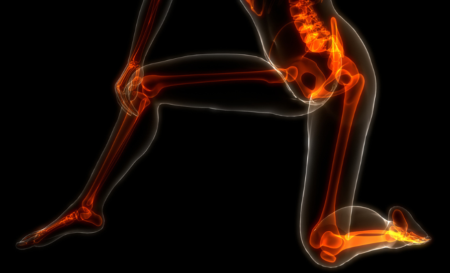
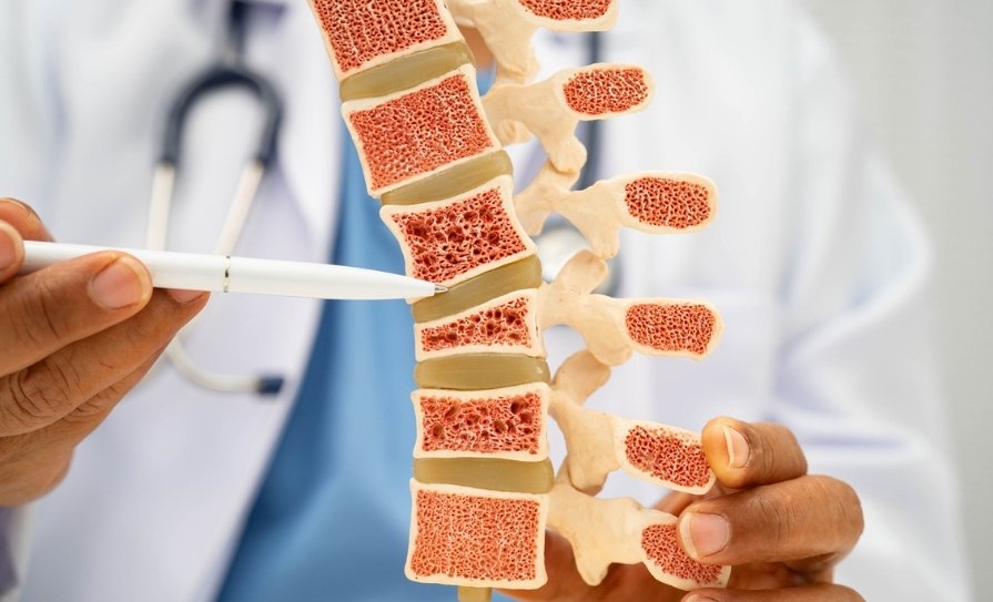
Osteoporosis is common and affects an estimated over 300,000 people in Ireland. Although more common in females who have gone through the menopause, it can also affect men and occasionally children. It is defined as a decline in bone mass associated with a microarchitectural deterioration in bone resulting in an increased risk of fragility fracture. In practise, the diagnosis can be made when the lowest T-score on DXA (dual energy x-ray absorptiometry) is -2.5 or below at any of three sites (total hip, neck of femur, or lumbar spine). However, a clinical diagnosis of osteoporosis can also be made without bone mineral density (BMD) measurements in patients who have had a fragility fracture of the hip or spine. Fragility fracture is one that occurs from a fall from standing height or due to trauma not normally expected to cause a fracture. Low bone mass or osteopaenia is defined as T-score between -1.0 to <-2.5. In pre-menopausal females and males aged under 50 years, a Z-score ≤-2.0 indicates low bone mass, which may be due to osteoporosis, though other causes such as osteomalacia need to be considered.
Which patients should have a DXA?
The International Society for Clinical Densitometry (ISCD) recommend a screening DXA in all females aged ≥65 and males aged ≥70. Post- or peri-menopausal woman under 65 and males under 70 years should also have a DXA if they have a risk factor(s) for low bone mass, eg, low BMI, prior fracture, smoking, medications, or a condition causing bone loss. DXA is also indicated where BMD needs to be monitored (eg, in those on treatment or to monitor bone loss due to drugs).
Who to treat?
Osteoporosis therapy may in general be initiated when there is: (1) clinical diagnosis due to low trauma hip or vertebral fracture; (2) diagnosis based on DXA criteria; (3) high fracture risk as calculated using tools such as FRAX (Fracture Risk Assessment Tool); and (4) osteopaenia with high-risk of fracture.
The FRAX tool can be used to estimate 10-year fracture risk with or without BMD. The threshold to start therapy varies by country and guidelines. The National Osteoporosis Guideline Group (NOGG) in the UK have established age-dependent thresholds. For example, in adults aged 70 and over, therapy is recommended when the 10-year fracture risk for hip is 5.4 per cent and 20.3 per cent for major osteoporotic fracture. In patients with no fracture history, but with moderate osteopaenia (T-score <1.5-2.0) and on drugs causing bone loss (eg, aromatase inhibitors, steroids), antiresorptive therapy can be started.
Factors to consider
Up to 50 per cent of fragility fractures occur in patients with osteopaenia. While about 70 per cent of bone strength relates to BMD, other factors collectively determining ‘bone quality’ are important, especially in the spine. For example, patients on steroids, aromatase inhibitors, and androgen deprivation therapy have a higher risk of fracture independent of BMD, resulting from their negative effects on bone.
The FRAX Tool
The FRAX tool is an osteoporosis risk assessment test, which uses information about bone density and other risk factors for breaking a bone to estimate a person’s 10-year fracture risk. A FRAX score estimates the chance of breaking a hip as well as the combined chance of breaking a hip or other major bones over the next 10 years. Other major bones include the spine, hip, forearm, and shoulder.
The FRAX tool can be used to guide treatment decisions in people who meet the following three conditions:
- Post-menopausal women or men aged 50 and older;
- People with low bone density (osteopenia);
- People who have not taken an osteoporosis medicine.
The FRAX tool is available at: https://frax.shef.ac.uk/FRAX/tool.aspx
| Risk Factor |
Description |
| 1. Age |
The risk of osteoporosis increases with age. |
| 2. Gender |
Women are more prone to osteoporosis, especially after menopause. |
| 3. Family history |
A family history of osteoporosis or fractures may increase the risk. |
| 4. Body weight |
Low body weight or being underweight is a risk factor. |
| 5. Ethnicity |
Caucasian and Asian individuals are at a higher risk. |
| 6. Hormone levels |
Low estrogen levels in women and low testosterone levels in men can contribute. |
| 7. Dietary factors |
Low calcium and vitamin D intake can impact bone health. |
| 8. Physical activity |
Lack of regular weight-bearing exercise may increase the risk, while overtraining can also increase the risk of developing osteoporosis. |
| 9. Smoking |
Tobacco use can weaken bones. |
| 10. Excessive alcohol consumption |
Heavy drinking can negatively affect bone health. |
| 11. Certain medications |
Long-term use of certain medications like glucocorticoids can increase the risk. |
| 12. Medical conditions |
Certain diseases, such as rheumatoid arthritis and digestive disorders, including coeliac disease or Crohn’s disease, may contribute to the development of osteoporosis. |
TABLE 1: Risk factors for osteoporosis
FRAX estimated fracture may be increased by 30 per cent in patients with recurrent falls (≥2 in last year). A recent fracture (within two years) is a big predictor of imminent fracture and needs to be considered when assessing fracture risk. Importantly, the majority (70 per cent) of vertebral fractures do not present clinically, but are associated with increased incident vertebral fracture risk by a factor of two-to-five. For this reason, any previous radiological imaging which includes the spine should be reviewed. A more significant degree of vertebral collapse is also suggestive of more severe osteoporosis. In patients at very high risk of fracture (FRAX risk 60 per cent above treatment threshold), T-score <-3.5, or recent vertebral fractures, parenteral or anabolic therapy should be considered.
Vitamin D/calcium and lifestyle
Lifestyle factors should be addressed in patients with osteoporosis, including avoidance of smoking and alcohol in moderation (<14 units per week) as this can lead to an improvement in BMD. Weight-bearing exercise (at least 30 minutes/day) is also advised. Additionally, resistive exercises to improve muscle strength and balance are recommended and where appropriate multidisciplinary input to reduce falls risk. After vertebral fractures, an exercise programme should be started as tolerated and tailored to the individual with emphasis on strengthening extensor spine muscles. However, physical activity that involves forward flexion of the spine should be avoided as should lifting heavy items, as both increase the risk of vertebral fracture.
Secondary causes of osteoporosis should be looked for, especially in younger patients or those with very low T-scores. In patients with vertebral fractures, serum electrophoresis should be tested to exclude the possibility of multiple myeloma. Parathyroid hormone level may also be assessed in patients with normal serum calcium to identify secondary hyperparathyroidism or the possibility of normocalcaemic primary hyperparathyroidism.
Most trials of osteoporosis medications have included patients on vitamin D/calcium supplements. However, not everyone needs supplemental calcium and the dose should be tailored to the individual. In general, total calcium intake should be ≥1,000mg combining diet and/or supplements. Patients should have a serum 25-hydroxyvitamin D level of at least 50nmol/l, which can usually be achieved with intake of 800-to-1,000IU of vitamin D per day. For this reason, all patients should have their vitamin D level checked with consideration for rechecking levels in those where compliance may be poor or with malabsorption syndromes.
Bisphosphonates
First-line therapy for osteoporosis is usually oral bisphosphonates, which are also indicated for prevention of steroid induced osteoporosis. They reduce the risk of fractures of the hip and spine by up 50 per cent and of the forearm by about 25 per cent. However, use is contraindicated in renal impairment (<35ml/min), gastro-oesophageal reflux disease (GORD), and oesophageal disorders (Barrett’s oesophagus and achalasia). Unfortunately, persistence with bisphosphonates is low at about 50 per cent at one year, highlighting the importance of selecting the appropriate patients for treatment: Avoid if unable to comply with instructions (sit upright for at least 30 minutes, full glass of water, no food) and if GI intolerance/malabsorption syndromes. Alendronate may have superiority over risedronate in BMD gains, but there is no evidence of any difference in anti-fracture efficacy. Ibandronate can be taken once monthly and may be an option for some patients with mild GORD.
Zoledronic acid is an alternative therapy when there are GI contraindications to oral bisphosphonates. It is also recommended as an initial treatment for patients in hospital with a hip fracture and can be considered in those at very high fracture risk. It is given as a once yearly intravenous infusion over 15-to-30 minutes. It has similar antifracture efficacy at the forearm and hip, but is superior at reducing vertebral fractures (about 70 per cent reduction). Standard therapy is for three years though in patients with severe osteoporosis and high-risk of fracture, it may be considered for up to six years. About 25 per cent of patients experience a mild acute phase reaction after the initial infusion (musculoskeletal pain and fever), which usually resolves within 72 hours and can be treated with simple analgesics.
Drug holidays, when and for how long?
Bisphosphonates have a long half-life in bone (10 years) with anti-resorptive and anti-fracture effects persisting for a period after therapy cessation. For this reason, patients may have a break from treatment or a ‘drug holiday’ though this needs to be closely monitored. Importantly, the concept of ‘drug holiday’ does not apply to other osteoporosis treatments where BMD drops after therapy cessation. Treatment with oral bisphosphonates is typically for five years and for three years with zoledronic acid, after which a ‘drug holiday’ should be considered.
Drug holidays may be appropriate in patients who after treatment have a T-score at the hip or spine of >-2.5, no recent fractures, and are at lower risk of future fracture. However, in older patients at high-risk of fracture (history of falls, previous hip or recent fractures) or on drugs causing bone loss, therapy may need to be continued (up to 10 years for alendronate and six years for zoledronic acid). Fracture risk can also be reassessed using FRAX and applying standard treatment thresholds.
Drug holidays are typically for 18-to-24 months with oral therapy and for up to two-to-three years with zoledronic acid followed by repeat BMD assessment. In patients where there is decline in BMD, or significant rise in bone markers, restarting osteoporosis treatment is usually appropriate.
Treatment failure
This may be considered to occur when there is a decline in BMD (greater than least significant change on DXA, which is up to about 5 per cent), no improvement in bone markers, or ≥2 fractures despite compliance with treatment. This should always prompt an investigation into potential reasons including drug malabsorption, undiagnosed secondary causes of bone loss (endocrine and medications), or poor adherence to lifestyle advice.
Denosumab
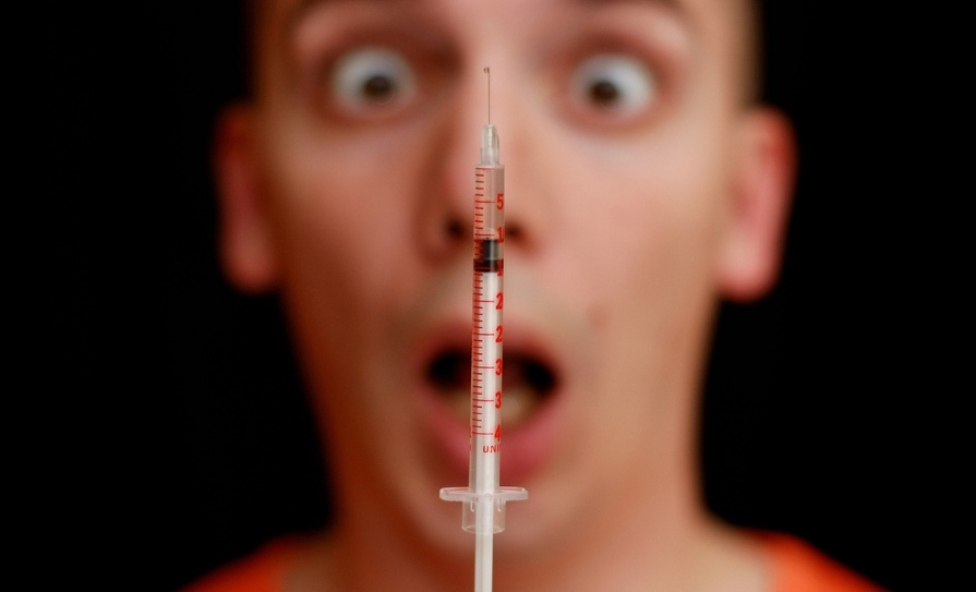
Denosumab is the most potent anti-resorptive used in the treatment of osteoporosis and is administered by subcutaneous injection twice yearly. It can also be used for the prevention of bone loss in patients on aromatase inhibitors or androgen deprivation therapy. It is a monoclonal antibody that blocks the action of the cytokine (RANKL), resulting in profound (up to 80 per cent) inhibition of bone resorption. It has a similar antifracture efficacy to zoledronic acid, but can be used in renal impairment (eGFR <30ml/min). Treatment for three years reduces fracture risk in spine by 68 per cent, 40 per cent at the hip, 20 per cent at non-vertebral sites, and 55 per cent at humerus. Therapy can be continued for up to 10 years (based on trial data), during which there is sustained rises in BMD and anti-fracture efficacy. Serum calcium should be normal and vitamin D >50nmol/l prior to starting so as to avoid hypocalcaemia. Denosumab is associated with an increased risk of cellulitis and possibly other infections and for this reason might not be as suitable for some patients.
As with many long-term medications, compliance can be an issue. Strategies to improve compliance include multi-component education, pharmacist support, shared decision-making, and treatment satisfaction. Recall systems to remind GPs and patients of when the next dose of denosumab is due are useful, as has been shown by research in Australia and other countries.
Rebound bone loss and fracture risk on stopping denosumab
Stopping denosumab results in rapid rebound bone loss, decline in BMD at both the spine and hip, and increased vertebral fracture risk (especially multiple vertebral fractures) occurring within months of discontinuation. In fact, all BMD gains from several years of treatment can be lost within 24 months with the greatest decline apparent in the first year. For this reason, denosumab must not be stopped unless patients are switched to alternative anti-resorptive therapies. The greatest predictor of bone loss after stopping denosumab is duration of use, while previous vertebral fracture strongly predicts incident vertebral fracture risk in the rebound period. While data on safety and efficacy beyond 10 years is lacking, some patients have now been on denosumab for 12 years (since it first became available) with no apparent increase in adverse effects. Importantly, the risk of atypical fractures is lower with denosumab than bisphosphonates.
For some patients already on denosumab and at high-risk of fracture (especially if recent vertebral fractures), remaining on treatment indefinitely or at least until T-scores improve (ie, ≥-2.5) and fracture risk reduces may be the best option. If stopping denosumab, recent NOGG guidelines suggest transitioning to zoledronic acid with close monitoring of bone turnover markers (BTM). The timing of further zoledronic acid therapy should be guided BTM. However, in patients at higher risk of fracture and when BTM are not accessible, a second infusion of zoledronic acid six months later is suggested. Alternatively, for patients, on denosumab for shorter periods (2.0-2.5 years), oral bisphosphonate (if no contraindication) after six months of their last injection can be considered with BTM monitoring to ensure adequate suppression of any rebound bone loss. However, despite follow-up therapy, there is still a risk of bone loss for some patients. For these reasons, in younger patients and in those with mild osteoporosis, starting denosumab may not be a good option. Conversely, in older patients with severe osteoporosis and/or life expectancy of up to 10 years, it is a good choice.
Teriparatide
Teriparatide is recombinant parathyroid hormone administered as a daily subcutaneous injection. It is used as a first-line therapy in patients with severe osteoporosis of the spine and/or low trauma or spontaneous vertebral fracture(s). It is the only anabolic therapy available in Ireland and reduces vertebral fracture risk by abut 70-to-80 per cent. However, its effect on hip BMD is modest where it has not been shown to reduce fracture.
Contraindications include unexplained raised (alkaline phosphatase) ALP, Paget’s disease, prior radiotherapy, hypercalcaemia, hyperparathyroidism, haematological or active malignancy, and renal calculi. Most patients tolerate it well though adverse effects include nausea, dizziness, musculoskeletal pain, palpitations, and low mood.
For patients who have severe osteoporosis of both the spine and hip, bisphosphonate treatment first may be more appropriate followed by teriparatide. Alternatively, dual therapy with denosumab and teriparatide for two years may be considered in a specialised setting.
Of note, switching from denosumab to teriparatide is associated with a decline in BMD at the spine (at three-to-six months) and hip (at 12 months) and is not advisable in patients with severe hip or spine osteoporosis.
Importantly, teriparatide must be followed up with further treatment (typically bisphosphonates) to consolidate BMD gains. For patients who have vertebral fractures despite denosumab therapy, the addition of teriparatide could also be considered in a specialised setting.
Atypical fractures
Atypical femoral fractures (AFF) are a rare and possible adverse effect of bisphosphonates (incidence of 1/1,000 after 10 years of oral therapy). They are a type of stress fracture of the subtrochanteric femur. Though rare, the risk increases substantially after five years and is greater in Asians and patients on steroids. Fractures often occur spontaneously or with minimal trauma and may be bilateral. As there is often a prodrome of pain in the groin/ thigh, patients on bisphosphonates for five or more years should have a femur x-ray if clinically indicated. This can identify ‘incomplete AFF’, which is generally an indication for prophylactic femoral nailing to prevent fracture propagation. When AFF occurs, bisphosphonates should be stopped, with data suggesting that this substantially reduces future AFF risk. However, in those at high-risk of fragility fracture due to their osteoporosis, the optimal future treatment strategy is unclear. The incidence of AFF with denosumab is lower and the vast majority of cases have occurred in patients who had prior bisphosphonate therapy.
Medication-related osteonecrosis of the jaw
This is a rare occurrence in the treatment of osteoporosis and appears more common with potent anti-resorptives (denosumab and zoledronic acid). In the majority of cases, it is precipitated by dental surgery, particularly dental implants or extractions. However, poor dental hygiene, diabetes, steroids, and anti-angiogenic cancer drugs are risk factors. The incidence of medication-related osteonecrosis of the jaw (MRONJ) appears to be higher with denosumab than zoledronic acid. Before starting these therapies, it is advisable to have a dental check and plan any dental surgery if needed in advance.
When MRONJ occurs, bisphosphonates may be stopped, though this needs to be balanced against risk of fracture. However, stopping denosumab is not recommended. For patients on denosumab and at higher risk of MRONJ, some guidelines recommend elective dental surgery five-to-six months after their last injection followed by re-administration about four weeks later. However, there is no clear evidence that stopping bisphosphonates in advance of dental surgery reduces the risk of MRONJ.
Decisions regarding osteoporosis treatment in patients with MRONJ should be made by an expert in osteoporosis.
Osteoporosis in younger adults
The majority of cases of osteoporosis in younger adults are associated with secondary causes including drugs, premature menopause, amenorrhoea, anorexia, and endocrine disorders. Osteomalacia resulting from vitamin D and or low calcium intake will also result in low BMD and should not be mistaken for osteoporosis. In patients at very high-risk of fracture and where other causes have been addressed or treated, bisphosphonates may be cautiously considered. However, females of child-bearing age should be counselled on the potential risk of teratogenicity and to avoid pregnancy while on treatment.
Resources
A number of useful tools and educational resources about osteoporosis for both healthcare professionals and patients are available on the Irish Osteoporosis Society website at www.irishosteoporosis.ie.