


Pilot will assess the feasibility of community-based lung cancer screening for high-risk individuals using low-dose CT scans
The Beaumont RCSI Irish Cancer Society (ICS) Lung Health Check (LHC) pilot is a new and important research study. It will examine the feasibility of lung cancer screening of targeted high-risk individuals through low-dose computerised tomography (CT) using a community-based mobile scanning unit. The pilot study is funded largely by the Irish Cancer Society, with additional support from the EU4Health SOLACE (Strengthening the Screening of Lung Cancer in
Europe) consortium.
This clinical trial is run by a team in Beaumont Hospital and the RCSI and is led by group Principal Investigator Prof Jarushka Naidoo, Consultant Medical Oncologist/Professor of Medical Oncology, Beaumont Hospital/RCSI. The study will aim to recruit high-risk individuals from selected GP practices in the north Dublin/north-east region and will operate over two screening rounds, one year apart. The trial hopes to start recruiting and scanning participants in the summer of 2025.
Lung cancer in Ireland
Lung cancer is the biggest cancer killer in Ireland and is responsible for 20 per cent of all cancer deaths in the country. This equates to over 2,000 deaths per year. The primary reason for this high rate of mortality is that patients present to lung cancer clinics often when they are symptomatic. This generally occurs when the disease is already at an advanced stage, at which point treatment options – though improving greatly – are less effective. Approximately 70 per cent of patients present to specialist rapid access lung clinics with locally-advanced or metastatic disease. Furthermore, roughly 25 per cent of all lung cancer patients have their first presentation through emergency departments with symptomatic disease.
Lung cancer, at its earliest stages, is asymptomatic. When detected at this stage, it can be effectively cured with surgery in the vast majority of cases, or with radiation therapy when surgery is unsuitable. The five-year survival rates for early-stage lung cancer are now routinely greater than 90 per cent when identified and treated correctly. However, unfortunately, detection at early-stage disease is often when investigations are being carried out for other reasons, ie, as an incidental finding.
Lung cancer screening is based on shifting the focus from diagnosing patients with symptoms, to identifying and treating early-stage disease in high-risk, asymptomatic individuals.
The lung cancer screening landscape
Lung cancer screening as a concept has been in existence since the 1970s, predominantly through chest x-rays and/or sputum cytology analysis. None of these studies, however, have ever shown a mortality benefit using chest x-ray as a screening tool.
The other major radiology modality that can investigate for lung cancer is CT. All CT scanners expose patients to small amounts of radiation.
Diagnostic CT scanners generally expose patients to about 7mSv (millisieverts) of radiation per scan. In lung cancer screening populations, scans need to occur over multiple rounds, potentially requiring years of imaging. As such, carrying out lung cancer screening with radiation exposure of 7mSv over multiple rounds offers an unacceptably high risk of radiation exposure, which hindered the development of CT as a lung cancer screening modality.
However, significant developments in CT technology have led to the development of low-dose and ultra-low dose CT scanners that significantly reduce the level of exposure. Low-dose CT scanners can now expose patients to as little as 1.5 mSv of radiation (the equivalent of a medium-distance airplane journey). These low-dose scans have been shown to easily identify pulmonary nodules (small spots on the lungs that can develop into lung cancer) as small as 4mm, making them potentially very useful for screening populations. The estimated risk of developing a cancer from 10 years of annual screening with a low-dose CT scan is less than 0.05 per 1,000 patients scanned.
The advent of low-dose CT scanners led to the development and publication of two seminal lung cancer screening trials in the New England Journal of Medicine (NEJM) over the past 15 years. The first was in the US – the National Lung Cancer Screening Trial – and was published in 2011. This trial of almost 55,000 high-risk patients (30 pack-year) compared annual CT scan versus chest x-ray (CXR) in high-risk patients aged 55-to-74 years, over three screening rounds one year apart, and then followed the patients for 3.5 years afterwards. The trial showed a reduction in lung cancer mortality of 20 per cent in the low-dose CT arm versus those who had CXR. This was followed by the NELSON trial in Europe, published in the NEJM in 2020, which followed 15,000 people over four screening rounds with a 10-year follow-up. This again showed a significant mortality reduction of 24 per cent in the CT arm after 10 years. Multiple other, smaller trials have all showed a similar trend in mortality benefit. As such, the evidence that lung cancer screening reduces morality in lung cancer is indisputable.
Despite this proven benefit, implementation of lung cancer screening programmes internationally has lagged. This is mainly due to poor uptake at population level for lung cancer screening in jurisdictions with functioning lung cancer screening programmes. According to the World Health Organisation, for screening programmes to be economically viable, uptake needs to reach 70 per cent of the target population. While established, mature, screening programmes in colon, breast, and cervical cancer routinely hit this target, lung cancer, though much newer, falls far behind. In the US, uptake of lung cancer screening as a percentage of the target population is often in single digits, and even in areas of highest uptake does not exceed 20 per cent. This makes the implementation of lung cancer screening challenging in more resource-limited settings.
Research attributes low uptake in lung cancer screening to a mix of socio-economic and psycho-social factors experienced by the people most at high risk for lung cancer. This results in physical and emotional barriers that inhibit screening uptake. Physical barriers in high-risk individuals are linked with those individuals from more socio-economically disadvantaged backgrounds where longer working hours, having more dependants at home, time constraints, and costs make attendance at hospital appointments difficult. Psycho-social barriers arise from the significant stigma associated with lung cancer. Particularly in the Western world, lung cancer is seen as a disease caused by lifestyle choice, and this has the effect of creating a sense of blame and fear for patients. This results in increased healthcare nihilism and a reluctance to engage with healthcare provision as a result. The phenomenon is backed up by data which suggests that current smokers are 50 per cent less likely to interact with screening services than former smokers.
The UK has been very proactive in overcoming these physical and emotional barriers to lung cancer screening through implementation of pilot studies over the last decade. Physical barriers have been overcome through the introduction of mobile-based scanning units, strategically placed in areas of high need with good public access. These include football
stadiums, shopping centres, and other community hubs.
Psychological barriers have been tackled in a number of effective ways. Firstly, placing GPs, who are patients’ most trusted healthcare providers, at the centre of the invitation process is key to improved participation. This, coupled with flexible appointment times, reminder letters, GP-based information leaflets, and other tools have proven to significantly increase uptake. Switching the focus from purely lung cancer screening to a more holistic review of lung health through encompassing symptom questionnaires, spirometry assessment for chronic obstructive pulmonary disease (COPD), non-judgemental smoking cessation advice, and other interventions can help remove some of the perceived stigma in lung cancer screening, while at the same time potentially initiating healthcare interventions that improve long-term morbidity and mortality.
These interventions have been incorporated in numerous UK-based screening pilots, which now routinely have screening attendance rates over 50 per cent, with many approaching 70 per cent. Lung cancer screening in the UK has produced data confirming cost-effectiveness ratios that make targeted lung cancer screening economically viable. As such, a national screening programme has been announced for NHS England to commence by 2029. This service will provide one million CT scans by 2029, with the identification of 9,000 extra lung cancers per annum, the vast majority of which will be treated with curative intent.
The Beaumont RCSI Irish Cancer Society Lung Health Check pilot
The European Union (EU) and the Irish Government have made lung cancer screening a policy priority, with recommendations for all EU countries to begin screening pilots to help inform EU policy. Recently, the ICS has partnered with Beaumont Hospital and the RCSI to announce a €4.9 million investment in lung cancer research. The Beaumont RCSI ICS Lung Outreach Programme, led by Prof Naidoo, is a multi-pillared partnership examining the areas of early detection, diagnosis, treatment, and survivorship in lung cancer. A key pillar of this programme is to investigate early detection through screening. The Beaumont RCSI ICS LHC will be Ireland’s first lung cancer screening pilot with the aim of assessing the feasibility of community-based screening, with mobile CT scanning units in the north Dublin, north-east area. The data gained from this study will be used to potentially inform policy on a future lung cancer screening service for Ireland.
The LHC pilot will encompass learnings from UK-based screening pilots and analyse their effectiveness in an Irish population and healthcare system. Key to this is the use of mobile CT scanning units in community locations. For the trial, a bespoke mobile CT scanning unit equipped with a state-of-the-art low-dose CT scanner and an adjacent mobile support unit with clinical rooms for LHC assessment has been commissioned, and will be operated by Alliance Medical diagnostic imaging. The unit will be staffed by nurses, radiographers, and administration staff for the duration of the pilot.
The mobile unit will operate in four main zones in the north Dublin/north-east region, with the unit based across four main sites, kindly provided by local GAA grounds. The support of these grounds, including Croke Park, has been pivotal to the ability to launch this pilot and it would not have been possible without their assistance. In addition to Croke Park, the grounds include Fingallians GAA club in Swords, O’Tooles GAA in Raheny, and O’Raghallaighs GAA club in Drogheda. The units will remain at these sites periodically, with study participants from surrounding, participating GP practices attending their LHC appointments at the venues.
Recruitment strategies are vital in the context of lung cancer screening. For this pilot study, GP-based invitation is a core tenet of the invitation methodology. As such, we have partnered with the Centric Health network of GP practices in the north Dublin/north-east region for the commencement of the trial. Our invitation methodology involves inviting all patients between the ages of 55-to-74 in participating Centric practices (19 in total) to take part. To do this, we will send an invitation letter to each patient, coupled with an information leaflet, explaining the study. The estimated Centric Health patient population aged 55-to-74 is approximately 30,000. Based on Health Research Board data, we anticipate that around 50 per cent will be ever-smokers, with a 30 per cent response rate and 40–to-50 per cent of respondents meeting high-risk criteria. From this, we aim to recruit just over 2,000 participants for an LHC.
In the invitation letter, each participant will be given a time and date for a telephone call with one of our study team, as well as an opt-out mechanism if they do not wish to take part. During that call, participants will be asked questions relating to inclusion and exclusion criteria before undergoing risk assessment using internationally validated risk-assessment scores, proven to outperform risk assessment with smoking pack year history alone. These include the prostate, lung, colorectal, and ovarian (PLCO) Score and the Liverpool Lung Project (LLPv3) risk-assessment scores, which are logistical regression models with multiple risk variables that include smoking history, family, and medical history, and other independent risk factors. Participants with a PLCO risk of ≥1.51 per cent and/or LLPv3 score ≥ 2.5 per cent on telephone assessment will then be invited for a formal LHC in the mobile unit at their nearest location.
Participants who attend an LHC will have a number of assessments on the mobile unit.
The first will be a respiratory assessment and baseline spirometry. High-risk patients for lung cancer screening are also high risk to have undiagnosed symptomatic airway obstruction (AO) suggestive of COPD. Similar screening cohorts have seen undiagnosed symptomatic AO rates of 20 per cent or more. Active case finding and identification of COPD and the initiation of earlier health interventions may help reduce overall morbidity and mortality in this high-risk cohort. Previously undiagnosed AO, identified in the course of the trial, will be referred to GPs, local community hub, or hospital services for appropriate intervention, depending on severity of airway obstruction.
There is growing evidence that using lung cancer screening for active promotion of smoking cessation can result in a 15 per cent increase in permanent smoking cessation rates, further improving long-term mortality in screening cohorts. For this study, we have worked closely with the HSE Tobacco Free Ireland programme to introduce a robust smoking cessation pathway whereby current smokers will receive a brief intervention on the mobile unit, and then will be referred to their local HSE smoking cessation services from the unit for further evidenced-based smoking interventions.
The pilot is based on similar, very successful UK-based screening pilots, which are now coalescing into a national screening programme in NHS England
An embedded translational biomarker study will involve selected participants providing blood, sputum, and breath samples. The aim is to investigate novel biomarker pathways in the context of lung cancer screening. In particular, the study will analyse biomarkers that could be used alongside screening radiology to enhance sensitivity and specificity in detecting lung cancer in a high-risk population.
All participants attending the mobile unit will then have a low-dose CT scan to complete their LHC. The scan itself only takes 45 seconds and can be carried out in a participant’s normal clothing. When the scan is completed, the LHC ends. The whole process should take less than 30 minutes.
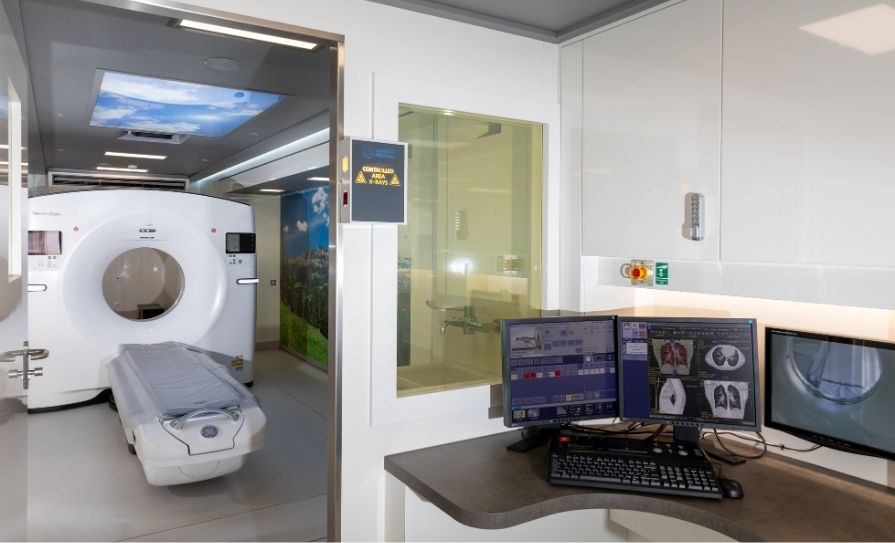
Credit: Susan Kennedy from Lensmen Photography and Video Production
Radiology pathway
All participants’ scans will be read by a team of thoracic radiologists, based primarily in Beaumont Hospital, who have undergone specific lung cancer screening training modules prior to the commencement of the trial. Each scan will also have an associated read by a dedicated artificial intelligence (AI) model (DeepHealth AI). This is specifically designed to provide volumetric analysis of pulmonary nodules. British Thoracic Society guidelines will be used in nodule assessment. All participants with no abnormal findings or small pulmonary nodules at their initial screening round scan (T0) will be invited back for a repeat CT scan in one year’s time in the mobile unit (T1). Participants with pulmonary nodules requiring closer surveillance will have a CT scan in the mobile unit at three months (T0+3). Participants with findings suspicious for lung cancer at any of these appointments will be referred directly by the study team to their nearest rapid access lung clinic (Beaumont Hospital or Our Lady of Lourdes [OLOL], Drogheda). The trial will end after the second screening round, with any participants requiring further CT surveillance at that stage being referred into the Beaumont Hospital or OLOL hospital network.
Significant incidental findings (IFs) are part and parcel of lung cancer screening. This is because the imaging can capture significant abnormalities in the lungs, great vessels, heart, and other surrounding organs. It is important to identify and manage participants with significant IFs, as these can have an effect on long-term morbidity and mortality. However, it is also critical to be careful not to over-report spurious findings that have little benefit and can increase unnecessary anxiety for participants. For this trial, a dedicated IF pathway protocol has been developed based on guidelines published by the American College of Radiology/NHS, and consensus guidelines published by the ERS. Furthermore, dedicated clinical referral pathways have been developed, whereby participants with significant IFs will be referred directly to RCSI hospital-based clinical specialists by the study team, or to their GP in other instances (specifically coronary artery calcification for primary prevention assessment).
Through a Beaumont Hospital virtual clinic, participants with a subset of significant IFs will be contacted by a member of the study team to discuss follow-up steps, as well as receiving written correspondence. Participants who have findings of nodules or IFs that are not detailed in the IF pathway or require further clinical discussion will have their cases discussed at a dedicated monthly lung health check screening multi-disciplinary meeting at Beaumont Hospital before finalising follow-up clinical plans. The LHC trial will use a dedicated lung cancer screening software tool (Nelson Plus) that will track all participants’ findings and produce templated letters that inform the participant themselves and their GP of the outcome of their LHC within 14 days of attending the mobile unit.
Further discussion
The Beaumont RCSI ICS LHC is a clinical trial that aims to assess the feasibility of carrying out targeted lung cancer screening using community-based mobile CT scanning unit. The pilot is based on similar, very successful UK-based screening pilots, which are now coalescing into a national screening programme in NHS England. The pilot study will run for at least two years. The data collected will be used to inform further discussions on the feasibility of a community-based lung cancer screening programme in Ireland.


Leave a Reply
You must be logged in to post a comment.