Reference: November 2025 | Issue 11 | Vol 11 | Page 26
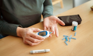
Diabetes Mellitus (DM) encompasses a group of metabolic disorders that are caused by multiple aetiologies and characterised by a state of chronic hyperglycaemia. If left untreated, DM can lead to widespread damage of organs and systems.
In addition to the better-known complications of DM like diabetic retinopathy, nephropathy, neuropathy, and vasculopathy, the burdensome disease also has a significant impact on bone strength, quality, and turnover, even when bone mineral density (BMD) appears normal.
Diabetic bone disease is a form of secondary osteoporosis that affects people with both types 1 and 2 DM. Patients with diabetes-induced bone disease exhibit variable degrees of bone loss and bone microarchitecture destruction.1 Ongoing disease results in a notably elevated risk of fracture and an impaired ability to heal post fracture of a bone.1,2
Osteoporosis, whether primary or secondary, is characterised by low bone mass and microarchitectural deterioration of bone tissue. It is usually diagnosed via dual-energy x-ray absorptiometry (DXA), and defined as a bone density of 2.5 standard deviations below the young healthy adult mean value (T-score ≤−2.5) or lower.
It is important to note that a DXA scan may underestimate fracture risk in diabetic patients as it does not take into consideration other DM-related issues such as changes in bone turnover, inflammation, and oxidative stress – all of which contribute to further weakening of the bone matrix.3 The FRAX tool also tends to underestimate fracture risk in people with DM.
This article looks at key aspects of DM, such as hyperglycaemia, insulin resistance, insulin-like growth factors (IGFs), advanced glycation end products (AGEs), and proinflammatory cytokines, that disrupt bone turnover by impairing osteoblast and osteoclast function, leading to imbalanced bone formation and resorption, and subsequently altered microarchitecture, strength, and quality.
Characteristics of diabetic bone disease
Increased fracture risk: Fracture is an under-recognised complication of DM that affects twice as many people with type 2 DM (T2DM), and up to seven times as many people with type 1 DM (T1DM), compared with their peers in the general population.4 Over one-third of T2DM patients exhibit bone loss and around 20 per cent meet diagnostic criteria for osteoporosis.1 The prevalence of secondary osteoporosis among patients with DM continues to grow considerably as incidence of the disease continues to rise across the globe.
Altered bone turnover: Stable bone remodelling – the cycle of bone formation by osteoblasts and resorption by osteoclasts – is crucial for maintaining bone strength and quality. However, in DM, this cycle is disrupted by a variety of mechanisms, particularly the hyperglycaemic environment. High circulating glucose stimulates osteocytes to produce sclerostin, a protein that inhibits bone formation by blocking the Wnt signaling pathway. Depression of this pathway leads to decreased osteoblastogenesis and bone turnover.3 Both T1DM and T2DM are associated with elevated serum levels of sclerostin. Interest is growing into the effects of anti-sclerostin antibodies on bone turnover.5,6
Impaired bone microarchitecture: Bone microarchitecture is also impacted through a variety of factors involved in the pathogenesis of diabetic bone disease – both molecular and structural – such as inflammation, oxidative stress, and microvascular complications, to name a few. Cortical bone accounts for 80 per cent of bone mass. In DM, there is often significantly reduced cortical thickness of the bone and weaker structure.
Trabecular Bone Score (TBS), which assesses the structure and quality of bone and predicts the risk of fracture, independent of BMD, is often reduced in people with DM. Reduced TBS and higher cortical porosity render the bone more fragile and prone to fractures.1 The TBS may be a better diagnostic tool than DXA alone.7
Pathogenesis
These hallmark characteristics of diabetic bone disease are, as mentioned, due to a variety of complex mechanisms that range from uncontrolled blood glucose levels to insulin deficiency and oxidative stress. The nature of DM drives many aspects of secondary osteoporosis development.
Insulin and IGF-1: Insulin and IGF-1 support healthy bone tissue and play a crucial role in osteoblast function. Insulin stimulates DNA synthesis, collagen formation, and osteocalcin production in osteoblasts, which are important precursors for bone tissue formation. Insulin also upregulates the expression of RUNX2, a gene essential for differentiation of osteoblasts and maturation of bone matrix.9
Additionally, IGF-1, which is structurally similar to insulin, is crucial for bone health as it supports the production of bone collagen and promotes bone matrix formation.1 People with DM often exhibit reduced levels of IGF-1.1,10
In patients with T1DM, the lack of insulin secretion and low IGF-1 negatively impacts bone mineralisation for a prolonged period of time. Since the onset of T1DM usually occurs during childhood/adolescence, these patients can experience a persistently low BMD throughout the lifespan.11
On the other hand, in the early stages of T2DM there is insulin resistance, leading to hyperinsulinaemia, and therefore, increased bone mineralisation. As T2DM progresses though, insulin secretion decreases.1 This process, alongside the other metabolic disturbances associated with DM, eventually leads to a reduced BMD and secondary osteoporosis in many patients.
Hyperglycaemia and accumulation of AGEs
Blood sugar control is a core element of care for the person with DM, and will also influence bone health. Research demonstrates that improved glycaemic control is associated with reduced, but not halted, bone loss.13 Studies show that osteoblast function and bone formation markers improve with intensive insulin therapy; however, resorption of bone remains elevated, possibly due to persistent inflammation.1,12
Hyperglycaemia leads to disruption of normal bone metabolism by inhibiting the activity of osteoblasts and osteocytes, reducing the synthesis of extracellular matrix components, and slowing down the mineralisation processes.
Moreover, the osteocyte lacuno-canalicular system formed by osteoblasts develops diabetes-induced microstructural disruptions. Hyperglycaemia also promotes the apoptosis and senescence of osteoblasts, further exacerbating bone loss. This process occurs in both T1DM and T2DM.1,14
A key component contributing to increased bone fragility in DM is the accumulation of AGEs, formed when elevated blood sugar levels lead to the non-enzymatic glycation of proteins such as collagen.1,15 Several studies have illustrated an association between poorly controlled blood sugar levels in DM and higher serum AGE levels, reduced bone strength, and increased risk of non-vertebral fractures, independent of BMD. Accumulation of AGEs in bone tissue has multiple effects, such as:
- Inhibition of osteoblast differentiation
- Impaired adhesion of osteoblasts to the collagen matrix
- Reduced expression of alkaline phosphatase, an enzyme that is critical for bone mineralisation
- Disruption of normal enzymatic cross-linking of collagen, leading to stiffening and weakening of fibres, and reduced elasticity and ability to absorb mechanical stress
- Reduced expression of pro-osteogenic markers like RUNX2 and Osterix
- Increased rate of apoptosis of osteoblasts
- Vascular effects like AGE-induced apoptosis of vascular smooth muscle cells, AGE-induced arterial calcification, reduced vascularisation and angiogenesis in bone. This leads to difficulty in supplying the bone and beginning the healing process following an injury.3,15,16
On the other hand, therapeutic glucose control is linked to reduced AGE accumulation and improved bone structure.15,16 Trials have shown that metformin provides a protective effect on bone quality as well as reduced AGE accumulation.1,3 AGE inhibitors have also demonstrated reduced bone brittleness and improved elasticity following 12 months of treatment.3
Pro-inflammatory cytokines and oxidative stress
In DM, prolonged inflammation, depicted by elevated levels of tumour necrosis factor-alpha (TNF-α) and interleukin-6 (IL-6), also markedly contributes to the development of diabetic bone disease. These cytokines promote activity of osteoclasts, leading to disruption of bone remodelling through a variety of pathways.1,3,17
TNF-α promotes the differentiation of osteoclast precursors into mature osteoclasts, leading to further bone breakdown, while IL-6 also promotes the differentiation of osteoclasts, leading to further overactivity of osteoclasts and ultimately excessive bone resorption. In addition, IL-6 promotes bone resorption through increased collagenase release and bone matrix degradation.1,17
DM induces oxidative stress through the formation of excessive reactive oxygen species (ROS), which damage various cell types, including bone marrow mesenchymal stem cells, which are critical for new bone formation.18 Signalling pathways involved in bone metabolism are also disrupted due to the circulating ROS and oxidative stress, leading to further bone loss.17,18
Research carried out on patients with T1DM showed elevated levels of TNF-α and IL-6, with elevated levels of these pro-inflammatory cytokines correlating with increased markers of bone resorption and reduced BMD.1 Studies investigating the effects of anti-TNF therapy and IL-6 inhibitors showed a reduction of osteoclast activity and bone resorption, and improvements in overall bone structure, despite an initial low bone mass.19 Anti-inflammatory drugs, metformin, and antioxidants have also exhibited positive effects in reducing ROS and oxidative stress.
Gut microbiota
The gut microbiome also appears to play a critical role in bone health, with ongoing research showing links between gut microbiota dysbiosis and DM.3,20 In healthy patients, short-chain fatty acids (SCFAs) such as butyrate, propionate, and acetate are produced by bacterial fermentation of dietary fibre in the colon. These SCFAs stimulate calcium absorption in the intestines and enhance osteoblastic activity, among other functions. Butyrate also functions as an anti-inflammatory agent counteracting the pro-inflammatory environment associated with DM.
Diabetes-related dysbiosis leads to reduced production of SCFAs in both T1DM and T2DM, increased intestinal permeability, and subsequent entry of endotoxins such as lipopolysaccharides to the bloodstream – further driving inflammation.20 Lipopolysaccharides stimulate additional TNF-α and IL-6 circulation, promoting further osteoclastic activity and bone resorption with reduced bone formation.21
The gut microbiota composition in DM patients has been found to have reduced numbers of beneficial bacteria including Bifidobacterium and Lactobacillus, in the dysbiotic guts of people with DM. Research illustrates a link between microbiota modulation – through high fibre diets and the use of probiotics – and higher levels of SFCA-producing bacteria, ultimately leading to improved calcium absorption, reduced inflammatory cytokines, and improved bone health.21
Management of diabetic bone disease
The cornerstone of diabetic bone disease management involves controlling hyperglycaemia through diet, lifestyle, and pharmacological agents (when appropriate) combined with anti-osteoporosis drugs. Bisphosphonates are the first-line drugs for most patients with DM and severe bone disease. In the elderly and patients with impaired renal function, denosumab may be preferred.
Conversely, sulfonylureas and thiazolidinediones are not recommended due to the risk to hypoglycaemia and the mechanism of action, respectively. The effects of anti-osteoporosis drugs on glucose metabolism, the individual patient’s needs, and hypoglycaemia-related falls risk should be considered. Avoiding hypoglycaemia is vital, as falls due to hypoglycaemia increase the patient’s fracture risk substantially.1,3
Patients should also be encouraged to engage in exercise and eat a diet rich in healthy whole foods that contain the nutrients essential for bone health, including protein, calcium, magnesium, and vitamin D. A Mediterranean diet based on fresh fruits, vegetables, and fish has been shown to reduce fracture risk and microvascular complications in people with T2DM.22
Patients should also be encouraged to eat adequate amounts of fibre and incorporate fermented foods into the diet. Maintaining a healthy weight is also important. Avoiding smoking due to its negative affects on bone health, and alcohol to limit falls risk, is also important.
An emerging area of research involves the role of gastrointestinal hormones on bone metabolism and remodelling, and subsequently, the effects of incretin-based therapies on bone health in people with DM. Glucagon-like peptide-1 (GLP-1) and glucose-dependent insulinotropic polypeptide (GIP) both promote bone formation and improved bone density in people with diabetes.3,23
Studies have shown that the combination of GLP-1 and GIP therapies improves overall bone health in DM patients, offering a dual benefit of managing DM and promoting bone strength.3,23 Moreover, this combination regimen is associated with enhanced fracture healing.3
Conclusion
Diabetic bone disease is a complex condition, driven by multiple interrelated mechanisms that disturb normal bone homeostasis, leading to abnormal bone turnover and poor bone mineralisation. Patients with T1DM and T2DM should have periodical bone health assessments as part of routine care. Management requires a multifaceted, individualised approach that encompasses pharmacological treatments, lifestyle and dietary advice, and fall prevention.
The data regarding the effects of new antidiabetic drugs, such as SGLT2 inhibitors and DPP4 inhibitors, are inconclusive. However, GLP-1 analogues show a positive effect on bone mineralisation. Interest is also centred in areas like the gut microbiome, incretin-based hormones, and antioxidant supplementation.
References
- Wu B, Fu Z, Wang X, et al. A narrative review of diabetic bone disease: Characteristics, pathogenesis, and treatment. Front Endocrinol (Lausanne). 2022;13:1052592. doi:10.3389/fendo.2022.1052592.
- Ferrari SL, Abrahamsen B, Napoli N, et al. Diagnosis and management of bone fragility in diabetes: An emerging challenge. Osteoporosis Int. 2018;29(12):2585-2596. doi:10.1007/s00198-018-4650-2.
- Sharma P, Sharma RK, Gaur K. Understanding the impact of diabetes on bone health: A clinical review. Metabol Open. 2024;24:100330. Published 2024 Nov 8. doi:10.1016/j.metop.2024.100330.
- Van Hulten V, Rasmussen N, Driessen JHM, et al. Fracture patterns in type 1 and type 2 diabetes mellitus: A narrative review of recent literature. Curr Osteoporos Rep. 2021;19(6):644-655. doi:10.1007/s11914-021-00715-6.
- Li Y, Luo Y, Huang D, Sclerostin as a new target of diabetes-induced osteoporosis. Front Endocrinol (Lausanne). 2024;15:1491066. Published 2024 Dec 10. doi:10.3389/fendo.2024.1491066.
- Meier C, Eastell R, Pierroz DD, et al. Biochemical markers of bone fragility in patients with diabetes. J Clin Endocrinol Metab. Published online May 8, 2023. doi:10.1210/clinem/dgad255.
- Rajan R, Cherian KE, Kapoor N, Paul TV. Trabecular bone score – An emerging tool in the management of osteoporosis. Indian J Endocrinol Metab. 2020;24(3):237-243. doi:10.4103/ijem.IJEM_147_20.
- Upadhyay P, Kumar S. Diabetes and bone health: A comprehensive review of impacts and mechanisms. Diabetes Metab Res Rev. 2025;41(5):e70062. doi:10.1002/dmrr.70062.
- Ouquerke A, Blulel J. Osteoblasts and insulin: An overview. J Biol Regul Homeost Agents. Published online December 14, 2020. Available at: https://pubmed.ncbi.nlm.nih.gov/33342206/.
- Biadgo B, Tamir W, Ambachew S. Insulin-like growth factor and its therapeutic potential for diabetes complications – Mechanisms and metabolic links: A review. Rev Diabet Stud. 2020;16(1):24-34. doi:10.1900/RDS.2020.16.24.
- Levran N, Shalev-Goldman E, Levy-Shraga Y. Bone health in children and adolescents with type 1 diabetes: Optimising bone accrual and preventing fractures. Nutrients. 2025;17(15):2400. Published 2025 Jul 23. doi:10.3390/nu17152400.
- Ravindran S, Wong SK, Mohamad NV, Chin KY. A review of the relationship between insulin and bone health. Biomedicines. 2025;13(6):1504. Published 2025 Jun 19. doi:10.3390/biomedicines13061504.
- Schwartz AV. Diabetes, bone, and glucose-lowering agents: Clinical outcomes. Diabetologia. 2017;60(7):1170-1179. doi:10.1007/s00125-017-4283-6.
- American Diabetes Association Professional Practice Committee. Classification and diagnosis of diabetes: Standards of medical care in diabetes – 2022. Diabetes Care. 2022;45(Suppl 1):S17-S38. doi:10.2337/dc22-S002.
- Kanazawa I, Sugimoto T. Diabetes mellitus-induced bone fragility. Intern Med. 2018;57(19):2773-2785. doi:10.2169/internalmedicine.0905-18.
- Khalid M, Petroianu G, Adem A. Advanced glycation end products and diabetes mellitus: Mechanisms and perspectives. Biomolecules. 2022;12(4):542. Published 2022 Apr 4. doi:10.3390/biom12040542.
- Ma X, Zhang X. Research progress of diabetic osteoporosis: A comprehensive review. Front Endocrinol (Lausanne). 2025;16:1595228. Published 2025 Sep 2. doi:10.3389/fendo.2025.1595228.
- Mangialardi G, Spinetti G, Reni C, Madeddu P. Reactive oxygen species adversely impacts bone marrow microenvironment in diabetes. Antioxid Redox Signal. 2014;21(11):1620-1633. doi:10.1089/ars.2014.5944.
- Huang Z, Xu Z, Wan R, et al. Associations between blood inflammatory markers and bone mineral density and strength in the femoral neck: Findings from the MIDUS II study. Sci Rep. 2023;13:10662. doi:10.1038/s41598-023-37377-6.
- Di Vincenzo F, Del Gaudio A, Petito V, et al. Gut microbiota, intestinal permeability, and systemic inflammation: A narrative review. Intern Emerg Med. 2024;19(2):275-293. doi:10.1007/s11739-023-03374-w.
- Lyu Z, Hu Y, Guo Y, et al. Modulation of bone remodeling by the gut microbiota: a new therapy for osteoporosis. Bone Res. 2023;11:31. doi:10.1038/s41413-023-00264-x.
- Esposito K, Maiorino MI, Bellastella G, et al. A journey into a Mediterranean diet and type 2 diabetes: A systematic review with meta-analyses. BMJ Open. 2015;5(8):e008222. doi: 10.1136/bmjopen-2015-008222.
- Liu H, Xiao H, Lin S, et al. Effect of gut hormones on bone metabolism and their possible mechanisms in the treatment of osteoporosis. Front Pharmacol. 2024;15:1372399. doi:10.3389/fphar.2024.1372399.