Reference: May 2025 | Issue 5 | Vol 11 | Page 40
Leg ulcers represent a significant clinical challenge globally, particularly among older populations. Chronic wounds not only impact the physical health of individuals, but also carry psychological, social, and economic consequences.1,2 In Ireland and across Europe, leg ulcers are a growing concern in community and acute healthcare settings, necessitating a multidisciplinary, evidence-based approach to care.
The European Wound Management Association (EWMA) and national guidelines, including those by the HSE in Ireland, advocate for best practices grounded in clinical research, expert consensus, and holistic patient management.1,2
Epidemiology and burden of disease
Leg ulcers, particularly venous leg ulcers (VLUs), are the most common type of chronic lower limb ulceration, accounting for 70-80 per cent of all leg ulcers. The prevalence of leg ulcers in developed countries ranges from 0.1 to 0.3 per cent of the general population, increasing to over 1 per cent in those aged over 65 years.3 In Ireland, community-based studies have found prevalence rates similar to European estimates.4
It is estimated that approximately one-quarter to one-half of acute hospital beds are occupied by patients with some form of wound. Among these, around 55 to 60 per cent are classified as non-healing wounds, which include infected surgical sites, pressure ulcers, and leg or foot ulcers.
It is also estimated that over 23 per cent of all hospital in-patients develop a pressure ulcer, with many cases occurring during their hospital stay for an acute illness or injury, indicating that a significant proportion of these ulcers are preventable.2
A 2019 review by O’Donnell et al, found that VLUs alone affect approximately one in 500 adults at any given time, with recurrence rates as high as 70 per cent within five years if not managed effectively.5 It is estimated that one in 50 people over the age of 80 has a VLU.2
The financial implications of leg ulcers are profound. In the UK, it is estimated that treating leg ulcers costs the NHS around £1 billion annually.6,7 While precise data on the cost burden of leg ulcers in Ireland is lacking, extrapolating from UK data and population size suggests an annual cost of €50-€100 million for the Irish healthcare system.6,7,8 Costs are associated with prolonged treatment duration, frequent clinic visits, nursing time, dressings, compression therapy, and complications such as infection or cellulitis.6,7,8
Wound care places a considerable financial burden on the healthcare system. Although there is no exact figure available for the cost specifically attributed to wound care within the HSE, it is estimated that the overall annual expenditure on wound-related healthcare amounts to approximately €788.5 million. On a per-patient basis, this equates to an estimated cost of around €3,850.2
Pathophysiology and classification
Leg ulcers are defined as wounds on the lower limb that show no tendency to heal after six weeks of appropriate care. They are typically classified by underlying aetiology into venous, arterial, mixed aetiology, diabetic, and pressure ulcers. Among these, venous ulcers are the most common, followed by arterial ulcers and those of mixed venous-arterial origin.9,10,11
VLUs result from chronic venous insufficiency (CVI), a condition where venous valves in the legs fail to function adequately, leading to venous hypertension. This causes capillary leakage, tissue oedema, inflammation, and eventually skin breakdown.
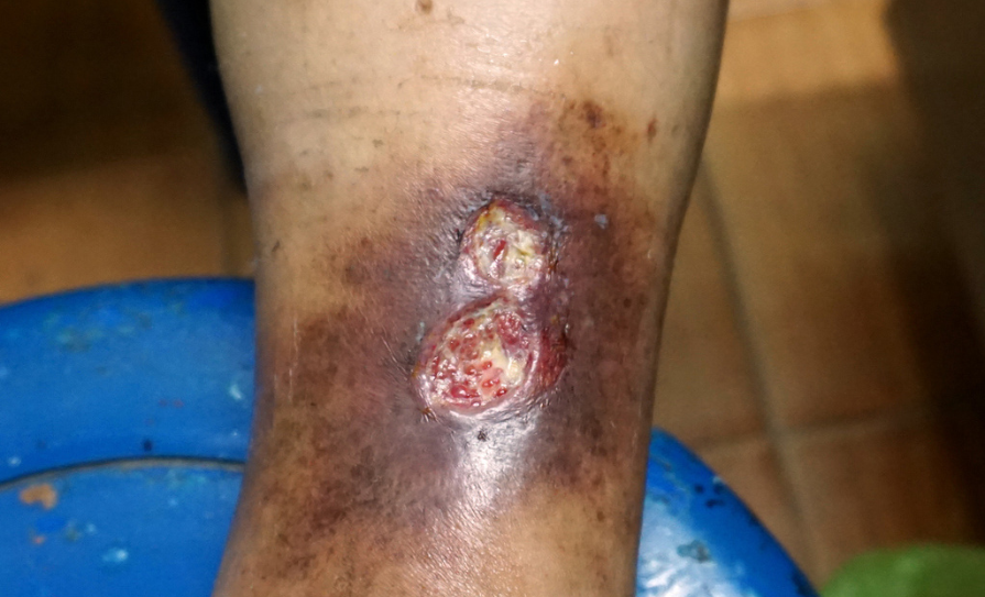
Clinical features include a shallow ulcer with irregular borders, often located in the medial lower leg (gaiter area), with associated signs of venous disease, such as varicose veins, lipodermatosclerosis, and haemosiderin staining.9,10,11,12
Arterial ulcers, in contrast, result from peripheral arterial disease, atherosclerosis, or thromboembolic events. These ulcers are typically painful, located over pressure points such as the lateral malleolus or dorsum of the foot, and have a ‘punched-out’ appearance with necrotic tissue.
Mixed ulcers exhibit features of both venous and arterial insufficiency and require careful management to avoid complications from inappropriate compression therapy.12,13
Diabetic foot ulcers, often neuropathic or neuro-ischaemic in origin, constitute a separate but overlapping category. Neuropathy, ischaemia, and infection interplay to cause ulcers, particularly over weight-bearing areas. Accurate assessment and classification are therefore essential to guide treatment.11,12,13
Assessment and diagnosis
Accurate diagnosis and assessment form the cornerstone of leg ulcer management. A thorough patient history is important, including duration and progression of the ulcer, comorbidities such as diabetes or vascular disease, lifestyle factors (eg, smoking, mobility), previous ulceration, and treatments tried. Clinical examination includes inspection of the ulcer’s location, size, depth, exudate, surrounding skin, and signs of infection.14
A key diagnostic tool is the ankle brachial pressure index (ABPI), used to assess arterial supply to the lower limb. This non-invasive test compares the systolic pressure at the ankle with that of the arm. An ABPI of 1.0-1.4 is normal, 0.8-1.0 is indicative of mild arterial disease (compression can generally be applied), 0.5-0.8 suggests moderate disease (requiring modified compression), and <0.5 suggests severe arterial insufficiency, requiring urgent vascular referral. Values >1.4 may indicate calcified, incompressible arteries, often found in diabetic patients, and necessitate toe pressure or Doppler waveform analysis.15,16
Duplex ultrasound scanning is important in diagnosing venous incompetence, particularly in cases of recurrent leg ulcers or when considering surgical intervention. This non-invasive imaging modality assesses both the anatomy and function of lower extremity veins, identifying issues such as venous reflux and obstruction. Its role extends beyond diagnosis, aiding in planning appropriate treatments, including surgical options.14
In addition to imaging, comprehensive patient evaluation for leg ulcers includes blood tests to screen for conditions like diabetes and anaemia, which can impede healing. Microbiological swabs are recommended to detect infections; however, antibiotic therapy should be guided by clinical signs and culture results to prevent overuse. If malignancy is suspected, a biopsy is warranted to obtain a definitive diagnosis.14
Differential diagnosis
While venous ulcers are the most prevalent form of chronic lower limb ulcers, it is important to consider other potential diagnoses, including arterial occlusive disease (either alone or in combination with venous disease), ulcers resulting from diabetic neuropathy, malignancy, pyoderma gangrenosum, and other inflammatory conditions.
In cases where chronic ulcers do not respond to vascular treatment, between 20-23 per cent may be attributed to conditions such as vasculitis, sickle cell disease, pyoderma gangrenosum, calciphylaxis, or autoimmune disorders.16
Treatment and management
Treatment options for venous ulcers include conservative management, mechanical modalities, medications, advanced wound therapy, and surgical options.16 Although the main goal of treatment is ulcer healing, secondary goals include reduction of oedema and prevention of recurrence. The overarching goal of treatment is to heal the ulcer, prevent recurrence, and manage underlying aetiology.14
Compression therapy is a key treatment for venous ulcers in patients without arterial disease, aimed at reducing oedema and pain, improving venous reflux, and promoting healing. It also helps prevent recurrence.16 Compression can be delivered using multi-layer bandage systems (eg, four-layer or two-layer systems), short-stretch bandages, or compression hosiery.14,16 Multilayer compression systems, especially elastic ones, are more effective than single-layer or nonelastic systems.
Barriers to its use include wound drainage, pain, difficulty with application, physical impairments, and leg deformities. Contraindications include significant arterial insufficiency and uncompensated heart failure.6
Elastic compression bandages adapt to the leg’s size and shape, offering sustained compression during rest and walking, with infrequent changes needed. Inelastic wraps provide high compression during ambulation, but are unsuitable for non-ambulatory patients or those with arterial compromise.
Compression stockings, with a recommended pressure of 20-30mmHg (preferably 30-40mmHg), can aid in ulcer healing and prevent recurrence. These should be replaced every six months, and for those struggling with application, aids like velcro stockings or donning devices may be helpful.2,14
Continued use of compression stockings is recommended after ulcer healing.16 The EWMA and HSE advocate graduated compression therapy of 30-40mmHg at the ankle for uncomplicated VLUs with an ABPI >0.8.1,2 Dressings should maintain a moist wound environment while managing exudate, protecting peri-wound skin, and preventing infection.
Intermittent pneumatic compression can be considered for patients with persistent, generalised oedema due to venous insufficiency, lymphatic obstruction, or significant lower limb ulceration. While it is more effective than no compression at all, its comparative effectiveness with other compression methods remains uncertain. However, when used alongside layered compression, intermittent pneumatic compression may enhance ulcer healing.16
No single dressing has been proven superior for VLU healing and choice should be based on wound location, size, depth, moisture balance, presence of infection, allergies, comfort, odour management, ease and frequency of dressing changes, cost-effectiveness, and availability.14,16
Regular wound cleansing, debridement of necrotic tissue (autolytic, enzymatic, mechanical, or surgical), and appropriate dressings are important supportive measures. Enzymatic debridement, particularly using collagenase, has been shown to effectively eliminate nonviable tissue, although there is no evidence to suggest it is superior to sharp debridement.
Larval therapy is another effective method that not only assists in debridement, but also has potential benefits such as disinfection, promoting healing, and inhibiting and eradicating biofilms. However, patient acceptance and the potential for pain are barriers to its widespread use. Autolytic debridement, which involves moisture-retentive dressings, is often used in conjunction with other debridement methods.16
Pain management is important. While many venous ulcers are not acutely painful, mixed or arterial ulcers can cause significant discomfort. Analgesics should be used in accordance with the World Health Organisation pain ladder, and neuropathic pain may require agents such as gabapentin or amitriptyline.14
Infection control involves identifying clinical signs, such as increased exudate, odour, erythema, or systemic symptoms. Topical antiseptics may be used for local infection, while systemic antibiotics are reserved for cellulitis or osteomyelitis. Chronic colonisation is not an indication for antibiotics.11,14
Advanced and adjunctive therapies
Venous ulcers refractory to therapy that do not improve within four weeks of standard wound care should prompt consideration of adjunctive treatment options. These include negative pressure wound therapy, skin grafting, biological dressings, and electrical stimulation. While evidence is still emerging, some therapies have shown benefit in specific cases.11,14,16
Surgical options for venous disease, such as endo-venous ablation, sclerotherapy, or vein stripping, can reduce recurrence rates.14,16,17 Recent European studies advocate for early surgical intervention in selected patients to promote faster healing and prevent relapse. The ESCHAR trial (UK) showed that combining compression therapy with surgical correction of venous reflux significantly reduced ulcer recurrence.17
For arterial ulcers, revascularisation (angioplasty, stenting, or bypass) may be required to improve perfusion and facilitate healing. Multidisciplinary team involvement, including vascular surgeons, tissue viability nurses, podiatrists, and wound care specialists, is essential for complex cases.11,13,14
Prevention and recurrence management
Preventing leg ulcers involves addressing the underlying causes such as venous insufficiency, arterial disease, and diabetes. Compression therapy is particularly important for individuals with venous insufficiency, as it helps improve blood flow and reduces the risk of ulcer recurrence. Maintaining good skin care by regularly moisturising the legs helps prevent dryness and skin breakdown. Physical activity, including walking and leg elevation, encourages better circulation and supports vascular health.1,2,14
Effective prevention also includes the management of comorbidities such as diabetes, hypertension, and cardiovascular disease. Adopting a healthy lifestyle by avoiding smoking, maintaining a healthy weight, and following a balanced diet further reduces the risk of ulcer development.
Regular monitoring for early signs of skin changes, swelling, or varicose veins allows for timely intervention before ulcers can form. Education and continued support play a vital role in helping patients maintain leg health and prevent recurrence.2,14
Recurrence of leg ulcers remains a major challenge, with up to 70 per cent of VLUs recurring within three to five years. Prevention strategies include continued use of compression hosiery, patient education, lifestyle modification, and prompt treatment of new skin breaks.11,14
The HSE recommends Class II (20-30mmHg) or Class III (30-40mmHg) graduated compression stockings as maintenance therapy after ulcer healing. Regular follow-up, nutritional optimisation, smoking cessation, weight management, and promotion of mobility are also integral.2,10 Structured community-based leg ulcer clinics and integrated care pathways, where available, improve outcomes and reduce unnecessary hospitalisation.
Psychosocial and quality of life considerations
Chronic leg ulcers significantly impact patients’ quality of life. They are associated with reduced mobility, social isolation, depression, sleep disturbances, and stigma. Studies across Europe, including Irish research, have highlighted the psychological toll of non-healing ulcers, particularly in older adults living alone.18 Comprehensive care must include psychosocial assessment, patient involvement in treatment decisions, and support services where needed.
Patients frequently report dissatisfaction with inconsistent care and communication between primary and secondary services. Continuity of care, use of standardised care plans, and effective communication are important to improving satisfaction and outcomes. In this context, digital imaging and electronic health records are increasingly being used to track healing, improve documentation, and coordinate multidisciplinary input.14
Current Irish and European guidelines
The HSE’s national wound care guidelines align with EWMA and National Institute of Health and Care Excellence recommendations.1,2,19 Key points include early use of compression for VLUs, thorough vascular assessment prior to compression, appropriate referral to vascular services, use of evidence-based dressings, and a multidisciplinary approach. European guidelines emphasise stratified care based on ulcer aetiology, prompt assessment, and consideration of health system structures.1,2,19
Leg Ulcer Centre Ireland
The Leg Ulcer Centre Ireland (LUCI) is a pioneering nurse-led service, established as a joint initiative between a Registered Advanced Nurse Practitioner (RANP) in tissue viability and a consultant vascular surgeon. Developed in response to fragmented, inefficient care for VLU patients in the Northwest of Ireland, LUCI implements the principles of Sláintecare, aiming to provide timely, integrated, and patient-centred wound care across both acute and community settings.20
The service adopts a hub-and-spoke model, where the vascular ‘hub’ delivers specialist diagnosis and same-day interventions such as duplex ultrasound and endo-venous ablation, while the community ‘spokes’ provide follow-up and coordinated care. Patients referred to LUCI are seen within three weeks and benefit from a ‘one-stop, see-and-treat’ clinic, significantly reducing delays, hospital admissions, and associated costs. In 2022, LUCI managed care for 1,797 patients, predominantly older adults.20
The RANP leads the delivery of a full care pathway, from diagnosis to treatment and follow-up, and ensures care continuity by collaborating with interdisciplinary teams including public health nurses, clinical nurse specialists, plastic surgery, geriatric, and radiology services. The initiative has demonstrated improved healing rates, reduced recurrence, enhanced patient satisfaction, and greater efficiency.20
Conclusion
Leg ulcers present a complex and growing healthcare challenge, particularly among ageing populations in Ireland and across Europe. Their multifactorial nature demands accurate assessment, timely diagnosis, and personalised management rooted in evidence-based practice.
VLUs remain the most common and burdensome type, with high recurrence rates and significant physical, psychological, and economic impacts. Best practice guidelines from the HSE and EWMA highlight the importance of early intervention, compression therapy, appropriate dressing selection, and a multidisciplinary team approach.
Innovative models such as the LUCI clinic demonstrate the value of integrated pathways in improving healing outcomes, patient satisfaction, and healthcare efficiency. Preventive strategies, continuity of care, and ongoing patient education are important in reducing recurrence and supporting long-term leg health.
As the demand for wound care services continues to rise, strengthening community-based, patient-centred services and fostering interprofessional collaboration will be essential to delivering high-quality, sustainable care for individuals with leg ulcers.
References
- European Wound Management Association (EWMA). Management of patients with venous leg ulcers: Challenges and current best practice. EWMA Document; 2016. Available at: https://ewma.org/wp-content/uploads/2024/02/Management-of-patients-with-venous-leg-ulcers_FINAL_2016.pdf.
- Health Service Executive (HSE). National Wound Management Guidelines. Dublin: HSE; 2018. Available at: https://healthservice.hse.ie/filelibrary/onmsd/hse-national-wound-management-guidelines-2018.pdf.
- Probst S, Saini C, Gschwind G, et al. Prevalence and incidence of venous leg ulcers: A systematic review and meta-analysis. Int Wound J. 2023 Nov;20(9):3906–21.
- O’Brien JF, Grace PA, Perry IJ, Burke PE. Prevalence and aetiology of leg ulcers in Ireland. Ir J Med Sci. 2000 Apr–Jun;169(2):110–2.
- O’Donnell TF, Passman MA, Marston WA, et al. Management of venous leg ulcers: Clinical practice guidelines of the Society for Vascular Surgery and the American Venous Forum. J Vasc Surg Venous Lymphat Disord. 2019;7(5):638–89.
- Guest JF, Fuller GW, Vowden P. Venous leg ulcer management in clinical practice in the UK: Costs and outcomes. Int Wound J. 2018;15(1):29-37.
- Posnett J, Franks PJ. The burden of chronic wounds in the UK. Nurs Times. 2008;104(3):44-5.
- O’Connor T, O’Loughlin A, McIntosh C, Cotter P, Murphy M, O’Connor M. Community leg ulcer clinics in Ireland: A cost-effective and patient-centred approach to care. J Wound Care. 2017;26(9): S30–S35.
- Gethin G, Probst S, Stryja J, Christiansen N, Price P. Evidence for person-centred care in chronic wound care: A systematic review and recommendations for practice. J Wound Care. 2020 Sep 1;29(Sup9b): S1-S22. doi: 10.12968/jowc.2020.29.Sup9b.S1.
- Health Service Executive (2024). Leg Ulcers- Overview. HSE. Available at: www2.hse.ie/conditions/venous-leg-ulcer/.
- Robles-Tenorio A, Ocampo-Candiani J. Venous Leg Ulcer. [Updated 2022 Sep 18]. In: StatPearls [Internet]. Treasure Island: StatPearls Publishing; 2025 Jan. Available at: www.ncbi.nlm.nih.gov/books/NBK567802/.
- Kolluri R, Lugli M, Villalba L, et al. An estimate of the economic burden of venous leg ulcers associated with deep venous disease. Vasc Med. 2022; 27:63–72.
- Mayrovitz HN, Wong S, Mancuso C. Venous, arterial, and neuropathic leg ulcers with emphasis on the geriatric population. Cureus. 2023 Apr 25;15(4): e38123.
- Atkin L, Robinson H, Phillips K. Leg ulcer management: Enabling evidence-based practice. Wounds UK. 2024 Sep 23;20(3).
- Cain M, Ousey K, Atkin L. Use of ankle-brachial pressure index to assess patient suitability for lower limb compression. Br J Nurs. 2022 Nov 10;31(20): S6-S14.
- Bonkemeyer Millan S, Gan R, Townsend PE. Venous ulcers: Diagnosis and treatment. Am Fam Physician. 2019;100(5):298-305.
- Gohel MS, Heatley F, Liu X, Bradbury A, Bulbulia R, Cullum N, Epstein DM, et al. A randomised trial of early endovenous ablation in venous ulceration. N Engl J Med. 2018 Apr 24;378(22):2105-2114.
- Janke TM, Kozon V, Barysch M, et al. How does a chronic wound change a patient’s social life? A European survey on social support and social participation. Int Wound J. 2023;20(10):4138-4150.
- National Institute for Health and Care Excellence (NICE). Leg ulcer infection: antimicrobial prescribing. NICE guideline [NG152]. Published 11 February 2020. Available at: www.nice.org.uk/guidance/ng152.
- O’Shaughnessy M, Kent S. Advancing nursing practice in Ireland: A pathway of care for nurse-led integrated venous leg ulcer management. J Vasc Nurs. 2024;42(2):110–4.