Reference: October 2025 | Issue 10 | Vol 11 | Page 14
Acute skin and soft tissue infections (SSTIs) are common bacterial infections. They are especially common in children. SSTIs present with a wide spectrum of severity from low-risk impetigo to life-threatening necrotising fasciitis.
Staphylococcus aureus (Staph aureus) is the most common pathogen in non-bullous and bullous impetigo. Purulent lesions (folliculitis, furuncles, and carbuncles) are usually caused by Staph aureus. Non-purulent infections (most cellulitis, erysipelas) are usually caused by Streptococcus pyogenes (Strep pyogenes).
SSTIs are classified according to the tissues involved – the superficial epidermis, (impetigo, ecthyma), the hair follicles (folliculitis, furuncle, carbuncle), and the deep fascia (fasciitis).
Staph aureus
Up to 40 per cent of the population are colonised with Staph aureus. Many patients presenting with a community-acquired SSTI do not, in fact, have pre-existing colonisation. Staph aureus has the potential to cause a broad spectrum of infection, ranging in severity from asymptomatic colonisation, to bacteraemia, osteomyelitis, pneumonia, or endocarditis.
When faced with an undifferentiated skin infection, the initial task is to check for the presence or absence of purulence, as pus usually indicates a Staph aureus infection. The prevalence of purulent SSTIs has increased since the emergence of community-acquired methicillin-resistant Staph aureus (CAMRSA). Staph aureus SSTIs have a high rate of recurrence.
Strep pyogenes
Group A β-haemolytic strep (Strep pyogenes) causes some cases of non-bullous impetigo. It is particularly unusual in children less than two years of age. Strep pyogenes may cause the rare but potentially fatal necrotising fasciitis. Nephritogenic strains of Strep pyogenes very rarely cause post-streptococcal glomerulonephritis.
Minor skin injury provides the portal of skin entry for Staph aureus. Trauma, lacerations, bites, scratches (eczema), burns, ulceration, chickenpox, and fungal infections may cause such injury.
Impetigo involves the superficial skin. The great majority of impetigo cases involve children. Exposed body areas such as the face and extremities are the most frequently involved sites. It is very contagious.
Non-bullous impetigo is the most common SSTI in primary care. While Strep pyogenes causes some cases of non-bullous impetigo, the majority are caused by Staph aureus. There is no conclusive evidence that treatment prevents nephritis. Acute rheumatic fever is not a sequela. Impetigo is not painful, but may cause a mild itch.
Non-bullous impetigo is characterised by 2-4mm erythematous macules that rapidly evolve to short-lived papules that in turn evolve to form thin-walled vesicle or pustules. These rupture, leaving a superficial erosion covered with a honey-coloured crust.
Bullous impetigo is characterised by vesicles and bullae (1-2cm in diameter), which are filled with clear yellow fluid that can become turbid or purulent. It tends to develop on clinically intact skin and intertriginous areas. Bullae rupture, leaving a thin, brownish crust. As lesions spread centrifugally, central clearing may give them an annular appearance. Lesions may coalesce to form large, reddish, superficial, round to oval erosions. A peripheral collarette of skin may be found at the edge of ruptured bullae.
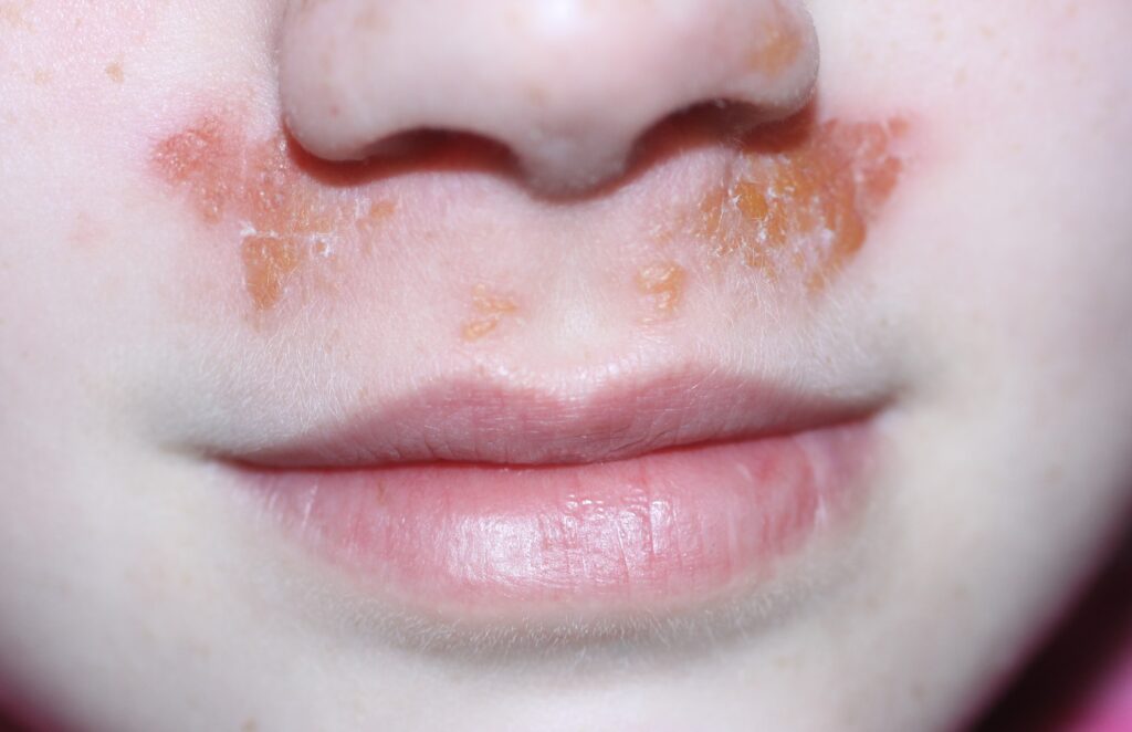
A: NON BULLOUS IMPETIGO
Characterised by 2-4mm erythematous macules that rapidly evolves to thin-walled vesicles (just under the patient’s nose) or pustules. Pustules rupture, leaving superficial erosions covered with a ‘honeycoloured’ crust.
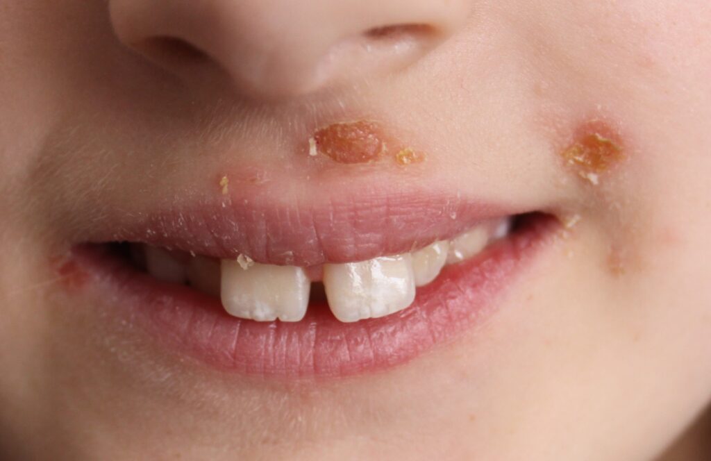
B: NON BULLOUS IMPETIGO
By the time patients present, the ‘honeycoloured’ crust may be the only finding. Note the area immediately adjacent to the upper lip demonstrating spontaneous clearing without treatment.
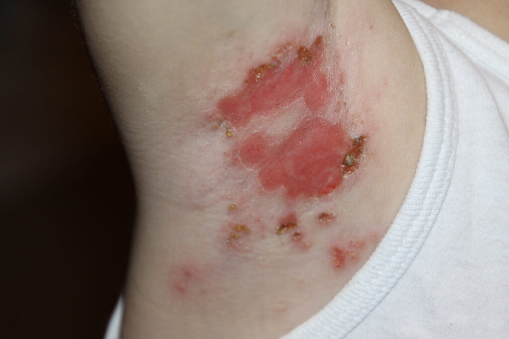
C: BULLOUS IMPETIGO
The fast advancing ‘honey-coloured’ crust periphery with central clearing. Vesicles and early bullae can be seen.
D: BULLOUS IMPETIGO
Is more likely to develop on clinically intact skin and intertriginous areas such as the axilla. Bullae rupture to give a brownish crust. Lesions may coalesce to form large, reddish, superficial, round to oval erosions. Some regard bullous impetigo as a localised form of staphylococcal scalded skin syndrome, as both are caused by similar toxin-producing strains of Staph aureus that cause severe blistering and exfoliation.
Management
Untreated impetigo resolves slowly, without scaring (two weeks for non-bullous impetigo, four to six weeks for bullous impetigo). Crusts can be gently removed by applying Vaseline, followed by cleaning with a warm, damp facecloth. As it is highly infectious, children should stay home from school until lesions have crusted over, or until two days after starting treatment.
NICE recommends hydrogen peroxide 1 per cent cream eight-hourly for five days for localised, non-bullous impetigo in patients who are not systemically unwell or at high risk of complications.
Advice on using a topical antibiotic is confusing, with some recommending a five-day course of fucidic acid. Because the risk of developing antibiotic resistance with topical antibiotics is higher than with the oral route, others advise a five-day course of oral flucloxacillin, if an antibiotic is indicated (eg, greater than three lesions). Oral flucloxacillin is recommended for ecthyma, although there is a lack of convincing evidence that this reduces scarring.
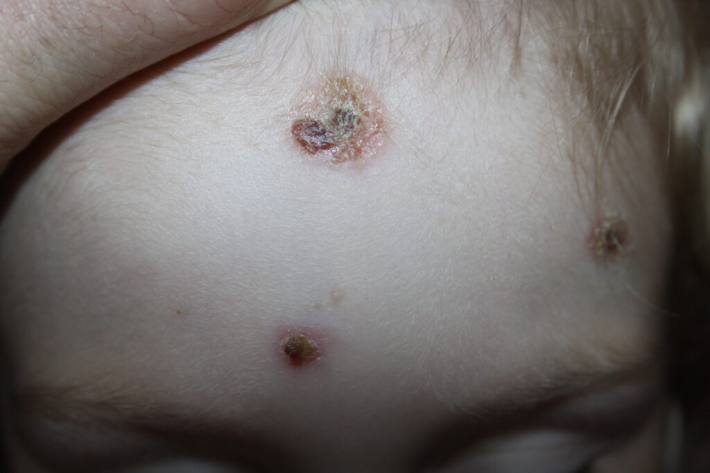
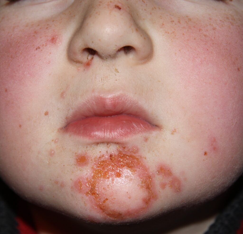
E: ECTHYMA
This may arise from impetigo, folliculitis, a scratch, an insect bite, or scabies. Initially puss accumulates under a gray-yellow scab. Progression into the dermis gives rise to a shallow ulcer, surrounded by erythema. If the scab is lifted, the sharply demarcated, evenly punched-out borders and the necrotic purulent base of the ulcer are seen. Crusts are frequently haemorrhagic. There is a significant risk of scarring.
Scalded skin syndrome
Like bullous impetigo, staphylococcal scalded skin syndrome (SSSS) is caused by exfoliative toxins that target desmoglein-1 in the skin. In infants, who have not yet formed antibodies to Staph aureus, these toxins disseminate throughout the body from the primary focus of infection, causing widespread erythema, flaccid blistering, desquamation, and erosions. Nikolsky’s sign is positive, with shearing of the epidermis from the dermis on lateral pressure.
SSSS favours intertriginous areas. It usually starts around the eye or the nasopharynx, so always check these sites for signs of blistering or crusting if SSSS is suspected. In slightly older children and adults, acquired immunity protects against SSSS, and bullous impetigo is a more likely presentation. SSSS can be life-threatening. Therefore, young children and immunocompromised patients require urgent admission.
Purulent SSTIs
Purulent SSTIs – folliculitis, furuncle, and carbuncle – present as localised collections of pus in cavities formed by necrosis and breakdown of the wall of the hair follicle. Folliculitis is an infection of the superficial portion of the hair follicle. Clinically it presents as clusters of papules and pustules on an erythematous base.
The scalp, extremities, paranasal area, axilla, and beard (sycosis barbae) are most frequently involved. With folliculitis, the inflammatory process is confined within the hair follicle whereas with a furuncle or carbuncle, the entire follicle and surrounding tissue is involved.
Between the dermis and underlying muscle and fascia, the hypodermis forms a layer of adipose tissue. Furuncles (also known as boils) develop from folliculitis, with infection spreading deeper into the hair follicle, producing a localised subcutaneous collection of pus within the hypodermis. A fibrinoid wall forms around the accumulated puss, separating it from the dermis and subcutaneous tissue.
A carbuncle is formed when several adjacent furuncles coalesce to form a cluster of involved hair follicles, connected by a sinus tract extending deeper into the hypodermis.
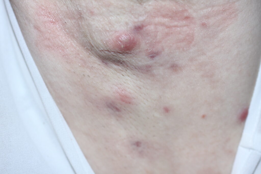
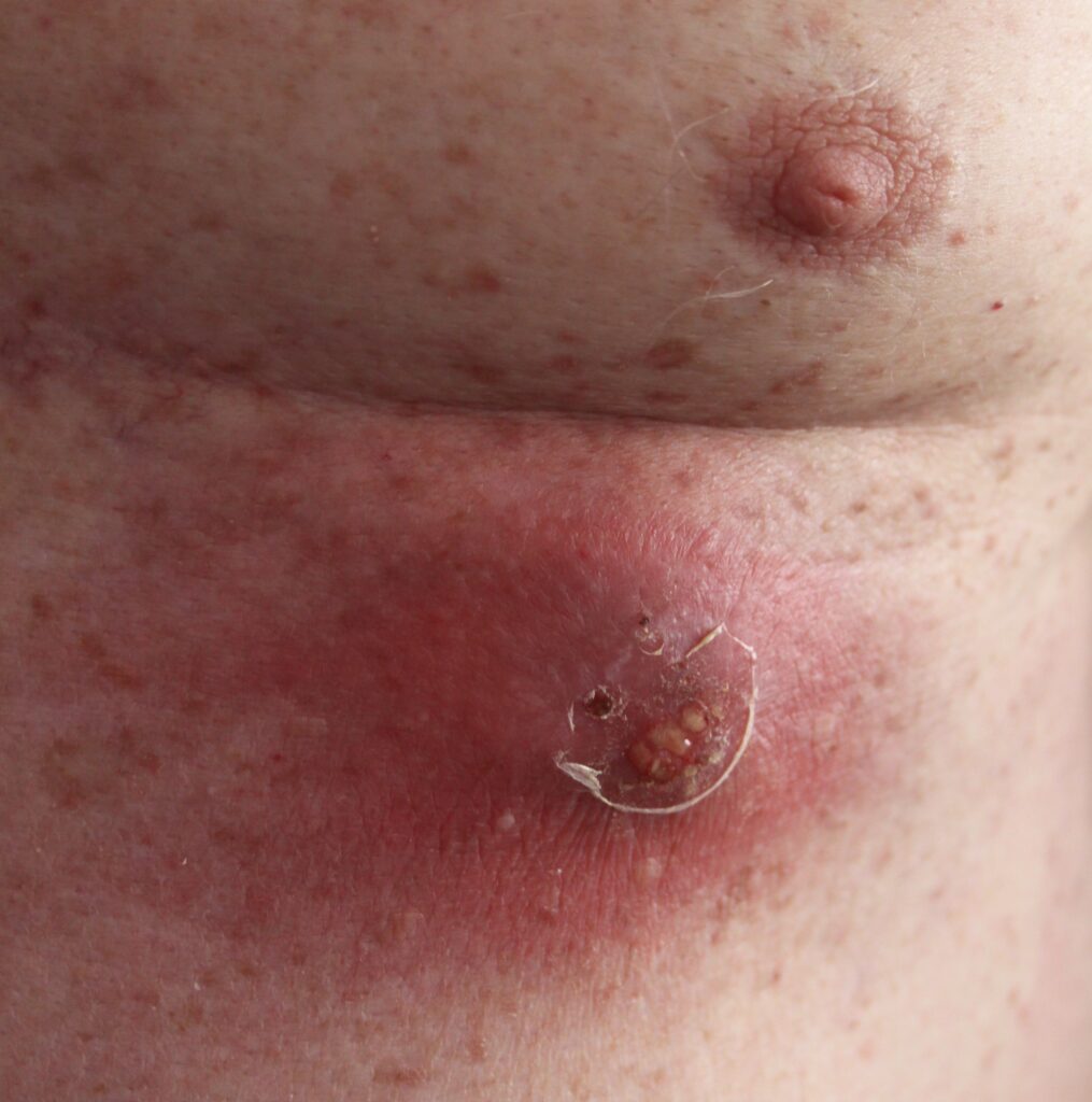
Multiple, recurrent furuncles in the axilla
This lesion from the chest wall of the same patient as slide F shows several adjacent furuncles coalescing to form a carbuncle with puss draining from three hair follicles
H: CARBUNCLE
A carbuncle on the lower chest wall, with surrounding erythema
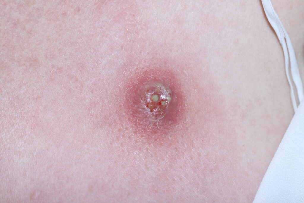
Clinical signs of furuncles and carbuncles include erythema, swelling, and induration, topped with necrotic skin or points of spontaneous drainage. Fluctuance is a classic finding, but may not be evident in early infection.
Cellulitis may surround the lesions, spreading radially. Pus drains from multiple drainage points on a carbuncle, representing the many adjoining hair follicles involved. Carbuncles are found most often on areas of thickened skin, such as the back, neck, and thigh.
Abscess denotes either a furuncle or a carbuncle. Not every abscess develops from a hair follicle infection, eg, breast abscess, perianal abscess. As it may be difficult to distinguish between folliculitis, furuncle and carbuncle, the term ‘purulent skin infection’ is increasingly used for any skin infection with pus.
The diagnosis is clinical, and swabbing for culture is rarely helpful. Applying moist heat promotes pus formation and drainage. Incision and drainage are recommended when fluctuance can be demonstrated.
Packing the drained cavity with gauze, following incision and drainage, is thought to be of no benefit.
Should an antibiotic be added to incision and drainage? Many large, placebo-controlled, randomised trials show benefit when an antibiotic is added, with an increase in clinical cure rates and reduced rates of recurrence. Many feel antibiotics are often unnecessary, especially when managing an uncomplicated furuncle or carbuncle. Some experts question the modest benefits in the above studies, as they may not outweigh the consequent increased risk of community antibiotic resistance. If an antibiotic is prescribed, choice is empiric and based on the clinical diagnosis.
There is no compelling evidence supporting superiority of any one antibiotic above another. As Staph aureus is the most likely pathogen, flucloxacillin is generally the first choice. Clindamycin is an alternative if the patient is allergic to penicillin. Clindamycin or doxycycline are recommended if MRSA is suspected.
Cellulitis
Cellulitis presents as an acute, spreading infection of the skin and subcutaneous tissues. The advancing borders are poorly defined (unlike erysipelas). Spread is sometimes patch-like. It is characterised by erythema, pain, and oedema, with local tenderness and warmth. Any break in the skin integrity is a risk factor (leg ulcer, trauma, tinea pedis).
Other conditions causing a red leg, such as lipodermatosclerosis and irritant contact dermatitis, are frequently misdiagnosed as cellulitis. Most cases of non-purulent cellulitis are caused by Strep pyogenes, with only a minority caused by Staph aureus. If pus is seen, the most likely cause is Staph aureus. It can be very difficult to find pus in cellulitis.
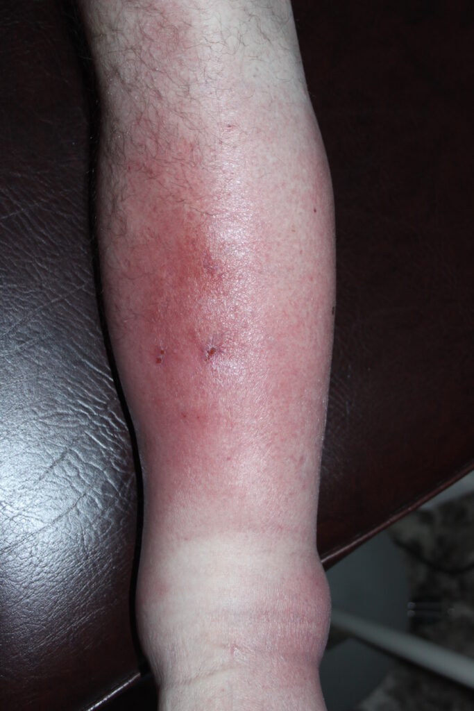
Note the non-distinct upper border
The patient had a history of axillary clearance. She recently suffered a minor prod of a thorn to her hand.
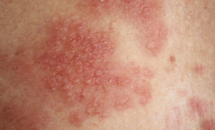
Erysipelas
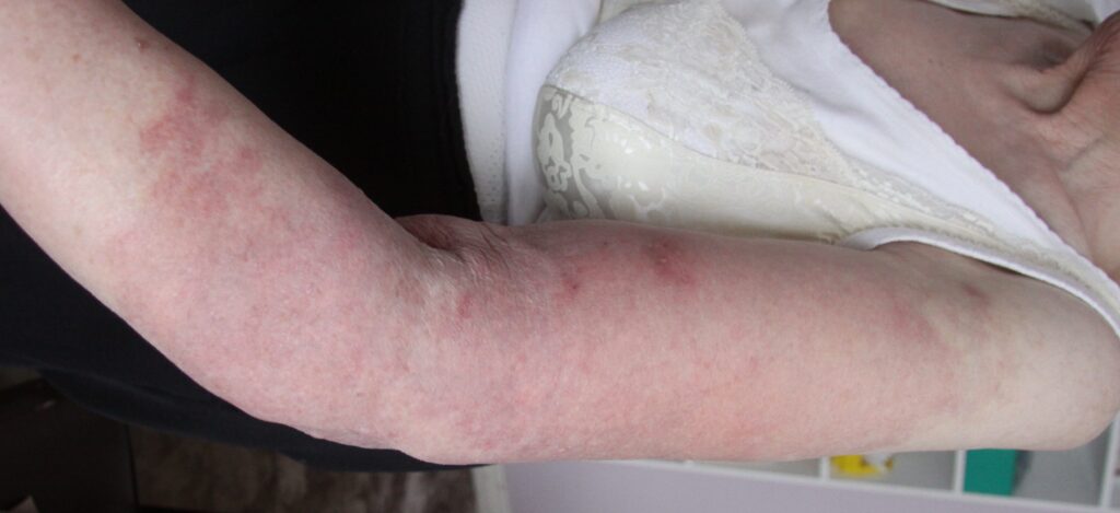
Presents as a painful, slightly raised, erythematous rash. Onset is acute, with a high temperature and vivid red colour. Unlike cellulitis, in erysipelas the inflammation is situated more superficially in the dermis. Many consider erysipelas to be a non-purulent form of cellulitis limited to the epidermis, and the terms are sometimes used interchangeably.
In erysipelas, borders are more sharply defined. It most commonly affects the face and the lower extremities. It spreads rapidly. Lymphoedema is a risk factor for erysipelas. The most common pathogen is Strep pyogenes. In more severe inflammation, serous, fluid-filled bullae may develop and should not be confused with pus.
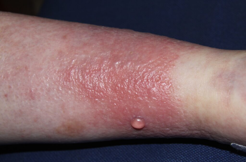
The intense inflammation has caused the development of bullae
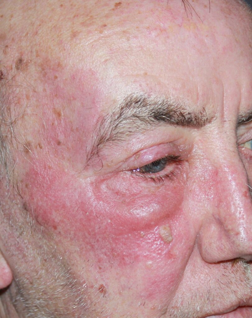
The rash is raised and has developed within the past 24 hours.
Management of cellulitis and erysipelas
In patients with cellulitis and erysipelas who show signs of systemic toxicity (fever, hypotension, tachycardia), are immunosuppressed, or have comorbidities (diabetes mellitus, heart failure, renal failure) should be admitted. Admission is advised for patients not responding promptly to oral therapy at home or if there is clinical deterioration at any time.
The majority are caused by Strep pyogenes. Therefore, β-lactam antibiotics are the first-choice antibiotic treatment. If Staph aureus is suspected, oral flucloxacillin 500mg QID should be taken for at least five days. MRSA coverage may be added if there is thought to be a risk.
Patients treated in the community should be followed up after 48 hours to check on the response to antibiotic therapy.
Necrotising fasciitis
Necrotising fasciitis is an infection of the fascia, a layer of connective tissue that lies below the hypodermis. Necrotising fasciitis often begins at the site of minor skin trauma, leading to rapid bacterial spread and necrosis of subcutaneous fat, muscle, and fascia. Fluid-filled bullae may develop. It spreads rapidly along fascial planes under the subcutaneous fat.
It is a life-threating infection. Urgent admission for prompt debridement is indicated. Watch out for signs of systemic toxicity (fever, chills, hypotension). Other signs include tenderness out of proportion to what you see, bullae or cutaneous necrosis with oedema, oozing a greyish fluid, crepitus, numbness, and rapid progression of symptoms and signs.
Classic clinical features are frequently absent at the time of presentation to primary care. Always keep the diagnosis in mind if a patient being treated for SSTI fails to respond to therapy, especially if their condition deteriorates. It is rare, but late diagnosis and delayed surgical debridement can be fatal.
- Severe or extensive disease
- Rapid disease progression
- Associated cellulitis
- Signs and symptoms of systemic illness
- Associated coexisting conditions or immunosuppression
- Very young or very advanced age
- Abscess in an area difficult to drain (eg, face, hands, or genitalia)
- Associated septic phlebitis
- Inadequate response to incision and drainage alone