Reference: May 2024 | Issue 5 | Vol 10 | Page 51
Hidradenitis suppurativa (HS), also known as Verneuil disease, acne inversa, or terminal hair dissecting folliculitis, is considered a chronic, progressive, relapsing inflammatory skin condition characterised by inflammation of hair follicles in multiple areas of the body, primarily intertriginous areas which contain apocrine glands.
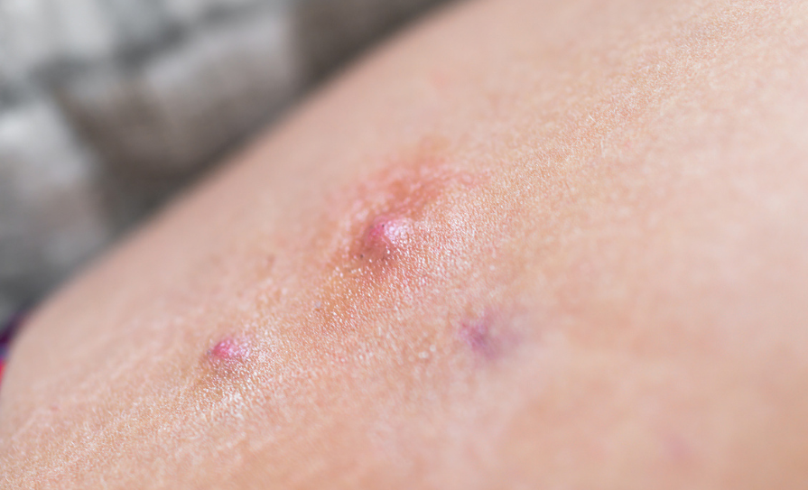
This manifests as painful nodules, abscesses, chronically draining fistulae, and subsequent scarring. The flexural areas most commonly affected include the axilla, inframammary area, gluteal, inguinal, perianal, and pubic regions.
Epidemiology
Global prevalence of HS has been estimated to be within the range of 0.00033-to-4.1 per cent. An approximate average delay of seven-to-10 years has been reported between onset of disease and diagnosis.
The prevalence of HS varies significantly depending on the geographical population and this is likely associated with different ethnicity dispersion among the different continents. With a female predominance, particularly in western countries, and typically occurring after puberty, HS has an average age of onset in the second or third decades of life.
Risk factors/causes
HS is a complex multifactorial disease. Evidence has suggested a link between HS and genetics, whereby 34 per cent of first-degree relatives have HS, according to previous research. The disorder is considered a complex polygenic disease in which a multitude of genetic variants play a role. Genetic research has detected various γ-secretase mutations involved in HS pathogenesis.
Numerous studies have also demonstrated a link between HS and obesity, with increased disease severity in people with higher body mass index (BMI). It has been suggested that obesity pathogenetically effects HS severity not only due to increased skinfold friction, but also as a result of low-grade systemic inflammation sustained by adipokine imbalance – augmenting the inflammatory processes in the skin of individuals with HS.
Another trigger factor associated with HS is cigarette smoking. The exact mechanism contributing to the pathogenesis of HS remains unclear, however, nicotine has been attributed to pathogenic events in HS such as follicular plugging, epidermal hyperplasia, neutrophil chemotaxis, keratinocytic cytokine production, and down-regulation of antimicrobial peptides (AMPs).
Smoking may also cause changes in the skin microbiome. In recent studies, patients with HS who actively smoke were noted to have a higher number of affected body areas than HS patients who do not smoke.
Mechanical irritation has been reported to be present before disease onset in some patients with HS. The use of antiperspirants is also believed to aggravate the disorder.
The role of sex hormones in HS
The skin is considered to be a steroidogenic machine due to its capability to produce most sex steroids de novo from cholesterol, and its ability to catalyse potent steroids from their respective precursors, playing a role in skin homeostasis.
Sex hormones, specifically androgen, progesterone, and prolactin, have been linked with HS. The female predisposition, peri- or post-pubertal onset, and frequent reports of premenstrual flare-ups in HS also indicates a role of sex hormones in HS.
Androgens can promote occlusion of the hair follicle as a result of increased proliferation of follicular keratinocytes, subsequently leading to follicular acanthosis, keratosis, and ultimately, plugging. Similarly, progesterone and prolactin may play a role in infundibular hyperkeratosis, follicular occlusion, and disease progression in HS.
Presentation
The diagnosis of HS is made on the basis of disease narrative, topography, and clinical features. According to the modified Dessau definition, there are three criteria to be met for the diagnosis of HS. These include: 1) the presence of typical lesions; 2) typical locations of lesions; and 3) chronicity.
Typical lesions of HS would include deep-seated painful nodules, bridged scars, suppurative sinus tunnels or tracts, abscesses, and double- and multi-ended comedones (also referred to as ‘tombstone comedones’).
As previously mentioned, the most frequently affected anatomical sites include axillary, inter- and infra-mammary, perineal, inguinal, perianal, and gluteal regions. Other less frequently affected sites include retroauricular, scalp, nape of neck, eyelids, lower abdomen, and suprapubic regions.
The hallmark of HS is chronicity and this is typically defined as two recurrences within a span of six months. The presence of these three criteria commonly provides high diagnostic specificity and sensitivity.
Disease course
Data on the long-term progression of HS is limited. On average, patients with HS develop a median of two lesions monthly, with every individual, painful boil lasting around approximately 6.9 days.
In general, the natural occurring frequency of flare-ups remains unidentified. However, triggers for flares have been observed and include diet, exercise, stress, friction, and weight gain. Furthermore, most patients describe local (eg, pruritus, erythema, and paraesthesia), or systemic (eg, headache, malaise, and fatigue) prodromal symptoms within 24 hours prior to development of a lesion.
Staging
There are various staging systems used for HS, with the two commonly used being the Hurley Staging System and Hidradenitis Suppurativa Clinical Response (HiSCR). The Hurley staging system, used in the clinical setting due to its simplicity and role in determining therapeutic needs, classifies the area which is affected the worst on the basis of the presence of single or multiple lesions and the presence of skin tunneling:
- Stage 1: Single or multiple abscess formation in the absence of sinus tracts or cicatrisation (wound healing for scar tissue formation).
- Stage 2: Single or multiple, recurrent abscess formation with tract formation and cicatrisation, widely separated lesions.
- Stage 3: Diffuse or almost diffuse involvement, or multiple interconnected tracts and abscesses spread throughout the entire area.
This staging system is convenient to assess baseline severity. However, due to the fact that it is based on scarring, unless patients get surgical intervention, they are likely to remain in their respective severity category.
The HiSCR is the most validated and commonly used tool for assessing response to treatment. It is a more recent staging tool that is based on counting the number of lesions. It is defined as a ≥50 per cent decrease in the number of inflammatory lesions (ie, inflammatory nodules and abscesses), and no increase in draining fistulae or abscesses when compared to baseline.
Other scoring systems used mostly in clinical trials include the Modified Sartorius Score (mSS), the Hidradenitis Suppurativa Severity (HSSI), the Hidradenitis Suppurativa Physician’s Global Assessment (HS-PGA), and the International Hidradenitis Suppurativa Severity Score System (IHS4), amongst others.
The microbiome in HS
Being the body’s largest communication with the external environment, the skin is the source of a multitude of commensal and symbiotic microorganisms. The skin microbiota is responsible for a number of essential homeostatic processes such as protection against pathogens, development of immune responses, and the breakdown of natural products via a metabolic exchange with host cells.
Differences in the microbiome have been observed between unaffected skin as well as lesional skin in patients with HS versus skin in healthy controls.
In HS, there is an alteration in the ecological structure of the skin microbiome. Anaerobic bacteria, such as Prevotella and Porphyromonas, coagulase negative Staphylococcus, and Staphylococcus aureus were the most frequently detected micro-organisms in HS lesions.
Both Prevotella and Porphyromonas in the skin microbiome may play a role in the pathogenesis of HS via upregulation of AMP secretion which subsequently enhances keratinocyte proliferation and recruitment of neutrophils and macrophages. Although the role of Staphylococci in HS is not clear, it has been demonstrated that such bacteria, which are known to lead to skin and soft tissue infections, are indeed found in lesions of HS.
Pathophysiology
1. The non-inflammatory stage
Initial skin changes in HS occur as a result of a complex interaction between genetic predisposition and environmental risk factors. In unaffected skin from predisposed sites, the infundibular outer root sheath exhibits hyperkeratosis and hyperplasia, which is likely a result of intrinsic dysregulation of the compartment of the hair follicle stem cell.
The outer root sheath shows replication stress provoking biogenesis of altered microRNA, type I interferon responses, increased secretion of chemokines (eg, RANTES [CCL5], IP-10 [CXCL10]), and upregulated expression of antimicrobial peptides (eg, S100A7-9, human β-defensin 2). These sub-clinical changes are supported by a shift in the skin microbiome towards a lower abundance of skin flora, a higher number of anaerobic bacteria, and a reduction in biofilm formation, collectively leading to a proinflammatory milieu.
There is the possibility that infundibular hyperplasia leads to follicular occlusion and stasis of bacteria and keratin within the dilating hair follicle. This cyst formation is accompanied by subclinical inflammation in the shape of a mixed mononuclear infiltrate positioned primarily around the follicle joined by small dispersed interfollicular sub-epidermal infiltrates.
Accompanied by mechanical stress, and possibly exposure to nicotine, this infiltrate promotes interfollicular hyperplasia and further influx of additional immune cells via the release of pro-inflammatory cytokines (eg, IL-7A, GM-CSF, and tumour necrosis factor (TNF).
2. The acute inflammatory stage
The breach or rupture of a cyst or tunnel exposes strongly immunogenic keratin fragments and bacteria to the dermis, which in turn causes an acute immune response characterised by a mixed immune infiltrate of myeloid cells (macrophages, neutrophils, and dendritic cells), followed by an influx and activation of B- and T-cells. The result of this process is clinically seen as inflamed nodules, abscesses, and draining tunnels.
After exposure to the contents of the sinus tracts and cysts, macrophages become activated and start producing various pro-inflammatory mediators such as IL-1ß, IL-8, and TNF. This, in turn, induces further expression of chemokines by keratinocytes and fibroblasts, which attract neutrophils, T-cells, and monocytes into the dermis during the process.
The response of fibroblasts to IL-1ß is the production of both additional chemokines and metalloproteinases (MMPs), such as MMP1 and MMP3 amongst others, which subsequently promotes cyst or tendril disintegration leading to non- or minimally-epithelial-lined tunnels.
Infiltrating monocytes become macrophages and dendritic cells which induce Th1 or Th17 cell development through production of either IL-12 and IL-23, respectively. Th1 cells, in turn, secrete IFN-γ, which causes activation of endothelial cells to aid immune cell infiltration and induces a positive feedback loop, which promotes additional development of the Th1 cell.
Th17 cells are prominent in lesional skin of HS and are responsible for the production of IL-22, TNF, and IL-17A, and amplification of the inflammatory cascade via the release of antimicrobial peptides and chemokines.
Local complement activation is outstanding in HS lesions and it promotes bacterial opsonisation, phagocytosis, and infiltration by neutrophils through production of C3a and C5a. The influx of neutrophils is further promoted via the production of additional chemoattractants by neutrophils, keratinocytes, macrophages, and fibroblasts.
Neutrophils in HS lesions exhibit increased formation of neutrophil extracellular trap (NET) and activation of the inflammasome NLRP3, which subsequently amplifies inflammation and activates plasmacytoid dendritic cells, leading to an augmented type I IFN (interferon) response.
Additionally, there is the release of elastase and proteinase-3, which are neutrophil serine proteinases, from the neutrophils onto NETs and these may cause proteolysis of inactive IL-36 precursor proteins into IL-36 isoforms which are highly active, further augmenting local inflammatory responses.
Plasma cells and B-cells have been described as having a key role in HR lesions and have been found in substantial amounts from early to chronic lesions. The latter tend to have the highest number of plasma cells, with a prominent increase in local immunoglobulins (Ig) production, which in turn form immune complexes within HS lesions.
Eosinophils and mast cells are also found within the lesional filtrate. However, they are less abundant than the other cell types. Eosinophils cause nerve ending stimulation and may play a role in HS-associated pruritus. Increased number of serum IgE levels helps with degranulation of mast cells within the lesion, causing the release of histamine and proteases. This process induces pruritus associated with HS.
Systemic effects may include fatigue and fever, with the likely cause of the latter being the production of endogenously-produced pyrogens including TNF, IL-6, and IL-1β.
3. The chronic inflammatory stage
As previously mentioned, single cyst ruptures along with associated flares on average last approximately seven days. Draining of inflamed tunnels or quiescence of acute lesions depends on clearance of the inflammatory infiltrate and is usually followed by a repair mechanism that aids with the restoration of the epithelial barrier between the cyst and/or tunnel lumen and the dermis of the skin.
Repeated occurrences of rupture of cysts and/or tunnels ultimately result in chronically inflamed lesions and these lesions typically show multinucleated giant cells commonly containing keratin fragments. Over-expression of B-cell activating factor (BAFF) and APRIL, which are B-cell survival factors, leads to the persistence of these cells and subsequent formation of tertiary lymphoid follicles consisting of B- and T-cells, follicular Th-cells, and follicular dendritic cells.
As lesions evolve, the number of B-cells increases with progressive differentiation towards plasma cells, memory B-cells, and plasmablasts, which are primarily found in end-stage lesions. Plasmablasts and plasma cells act as the primary source of augmented IgG expression in HS skin lesions, which in turn contributes to the formation of the local immune complex.
The infiltrate in chronic lesions appears to be structured in a way so as to contain the inflammation, with neutrophils located within the centre of the cyst breach surrounded by scattered lymphocytes and macrophages. Plasmablasts and plasma cells construct the outer layer of immune cells, which is subsequently encapsulated by a dense layer consisting of collagen and fibroblasts. The formation of tertiary lymphoid follicles typically occurs in the adjacent fibrotic dermis.
Additional architectural alterations, including fibrosis, are driven by activated fibroblasts and can be severe, resulting in lymphoedema, particularly in the pubic area, vulva, and scrotum. Any single collection of keratinocytes or remnants of cyst wall epithelium which remain in the dermis after cyst or tunnel destruction may be a potential source allowing for further formation of new tunnels, propagating the disease process even further.
Diagnosis
The diagnosis of HS is made primarily on the basis of the clinical picture. However, there are useful diagnostic tools which are helpful in the process. Ultrasound (US) imaging has been used in order to further characterise morphology and depth of lesions. For example, it is able to demonstrate subclinical fluid collections, augmented dermal thickness, and follicular dilatation in the early stages of the disease, and the formation of sinus tracts in the more advanced stages of the HS. Furthermore, the use of colour Doppler US may recognise subclinical sinus tracts, which is helpful in treating more precisely.
Histopathology studies tend to exhibit follicular epidermal hyperplasia and keratosis, infundibular lymphocytic infiltration accompanied by CD-8-cell epitheliotropism, sinus tracts, cysts, and tunnels in the absence of primary apocrine involvement in the early process of skin lesion formation. Biopsies of more advanced lesions may show follicular rupture and dermal fibrosis.
Additionally, the use of non-specific inflammatory markers such as erythrocyte sedimentation rate (ESR), C-reactive protein (CRP), and leukocytosis with neutrophilia may be used for the assessment of the systemic inflammatory burden caused by the disease. Research is taking place regarding the use of other proposed clinical biomarkers such as Chitinase protein 1, S100A8/A9, serum hepcidin, serum adiponectin, serum resistin, serum visfatin, and serum amyloid A, amongst many others.
Differential diagnosis
The differential diagnosis would include staphylococcal infections, abscesses, cutaneous Crohn’s disease, deep fungal infections, inguinal Hodgkin’s, lymphogranuloma venereum, and tuberculous lesions of the scrofuloderma type.
Management
There are several guidelines relating to the management of HS. Underlining all of them is the need for a multidisciplinary approach to the condition. The appropriate treatment of choice is predominantly determined by the clinical severity of HS.
Non-pharmacological interventions
- Smoking cessation is recommended as it has the potential to improve the severity and duration of the disease as well as other health-related outcomes.
- Weight reduction is also advised in view of the positive correlation between higher BMI and a more severe course of HS.
- Wound care and dressing should be customised based on the location of the lesion and the degree of drainage.
- Advice should be given to patients regarding wearing loose-fitting clothes so as to avoid friction and mechanical stress.
- Screening for anxiety and depression is also implemented across different guidelines. Psychological support may be required.
Pain management
- Topical analgesic options include lidocaine 5 per cent ointment or diclofenac 1 per cent gel.
- The use of NSAIDs (non-steroidal anti-inflammatory drugs) as systemic analgesics is recommended for acute pain management of HS.
- If reduction of severe HS-related pain is not achieved with first-line options, then the use of opioids such as codeine and oxycodone may be considered.
- Anti-convulsant drugs such as gabapentin and pregabalin, mostly in combination with NSAIDs, can adequately treat neuropathic pain associated with HS.
Topical therapy
This form of treatment is most useful in localised disease or as an adjunct to systemic therapies and is usually advised for milder stages of the disease. The use of topical antiseptics, such as chlorhexidine, and the use of topical antibiotics, such as clindamycin 1 per cent lotion, aim to decrease bacterial colonisation and related inflammation.
Usage of topical keratolytics, such as resorcinol 15 per cent cream, aim to decrease follicular clogging. Intralesional corticosteroid injections are used in individual acute flares and in cases of refractory nodules and sinus tracts. These injections can either be used alone or in conjunction with systemic treatments.
Systemic therapy
Oral antibiotics are the mainstay of treatment in HS. The options widely used in HS include oral tetracyclines (first-line), a combination of clindamycin and rifampicin (second-line), dapsone (third-line), or a combination of metronidazole, moxifloxacin, and rifampin (third-line). Intravenous ertapenem is reserved for severe cases of HS which are refractory to treatment.
Zinc salts, mainly zinc gluconate, exhibit anti-inflammatory effects in HS, likely due to modulation of cytokine expression, anti-androgenic properties, and inhibition of neutrophil granulocytes chemotaxis. Oral retinoids, such as acitretin, have a useful impact in HS since they cause a reduction in keratinocyte proliferation, hence resulting in the prevention of the plugging of the pilosebaceous unit.
Metformin also has a role in HS, not only due to its effect of insulin reduction, but also due to its antioxidative properties and ability to inhibit proliferation and inflammatory responses in keratinocytes. It is also known to reduce androgen production.
The use of combined oral contraceptives has frequently been recommended in the treatment of HS, particularly in a subset of female HS patients who experience a perimenstrual worsening of symptoms (up to 43 per cent of female patients with HS). Other anti-androgenic therapies include finasteride – a type II 5α-reductase inhibitor – and spironolactone – a potassium-sparing diuretic. Spironolactone also suppresses inflammation caused by TNF-α.
Biologics
Immunomodulation with biologics is gaining popularity in the management of more advanced forms of HS. Adalimumab, an anti-TNF-α drug, is the only internationally registered biologic approved for the treatment of HS. It is safe and effective, and is usually used for cases in which conventional treatments have failed. Infliximab, which is also an anti-TNF-α drug, has been recommended as a second-line biologic after adalimumab.
Surgical therapy
Surgical intervention should be considered in patients with severe chronic lesions which have either not responded to non-surgical therapies or else, are irreversible. Selection of the appropriate surgical therapy is based on the affected body region and the severity of the disease. Surgical procedures include radical surgical incision, deroofing, incision and drainage, laser therapy, wide local excision, and skin-tissue sparing incision with electrosurgical peeling (STEEP).
Association with other medical conditions and cancer
HS is associated with an array of diverse disorders. It is well-known for its contribution to the follicular occlusion triad which is characterised by the presence of HS, dissecting cellulitis of the scalp, and acne conglobata. This triad may develop into a tetrad if it occurs together with a pilonidal sinus.
Individuals with HS are at an increased risk of developing hyperlipidemia, hypertension, diabetes acromegaly, Cushing’s disease, metabolic syndrome, and polycystic ovarian syndrome. Other associated comorbidities with strong auto-inflammatory components include autoimmune conditions such as inflammatory bowel disease and psoriasis; syndromic comorbidities such as synovitis-acne-pustulosis-hyperostosis-osteitis syndrome; and rheumatological comorbidities such as the seronegative spondyloarthropathies.
Patients with HS appear to be at an overall increased likelihood of malignancy, with a 50 per cent increased risk, and are also 4.6 times more prone to the development of cutaneous squamous cell carcinoma (SCC). SCC is the main type of dermatological cancer reported in patients with HS, including those developing from long-standing inflammatory lesions such as Marjolin’s ulcers.
SCC occurs at a higher ratio in males than females and the most likely culprit for its development could be the human papillomavirus. Other specific cancers associated with HS include oral cavity and pharyngeal cancer, colorectal cancer, central nervous system cancer, and prostate cancer.
Paediatric HS
In spite of the fact that the disease commonly occurs after puberty, there have been cases described in children on the verge of puberty. This may be attributed to family history for HS. The disease may not actually be rare in children, but the low rate may be a result of delays in seeking care and the potential of under- or mis-diagnosing.
The paediatric population displays similar, but milder, clinical features and comorbidities as adults. Notable comorbidities in the paediatric population with HS include acne, hirsutism, and pilonidal cysts.
Impact of HS
In view of its chronic nature and regular relapses, HS has a considerable impact on patients’ quality-of-life (QoL) as well as on family members and caregivers. Among skin diseases, HS has one of the highest Dermatology Life Quality Index (DLQI) scores. The most outstanding component affecting QoL of such individuals is pain, and it is usually associated with the inflammatory nodules or abscesses.
Along the disease course, pain is reported by approximately 97 per cent of patients with HS. Pain, in turn, leads to poor sleep quality and insomnia. Another impacting factor on QoL is malodour.
Individuals with HS also often suffer from loneliness and social stigma, sexual impairments, and poor mental health including depression and anxiety. The condition also impacts the professional life of affected individuals, with an approximated working ability of 20 per cent lower than that of the average employee and an unemployment rate double that of the general population.
Conclusion
HS significantly affects all aspects of a patient’s life and often carries a high burden of treatment, therefore, psychological care is an important element of disease management. A multidisciplinary approach will ultimately improve patient outcomes, particularly in severe HS. Improved understanding of the pathophysiology, as well as novel biological drugs and other treatment strategies are providing relief for many patients, and research in the area is ongoing.
References on request