







Reference: May 2026 | Issue 5 | Vol 12 | Page 11
Pruritus of the genital and perianal skin is common but is probably underreported due to patient embarrassment. The pruritus is often intense and may lead to considerable physical and psychosocial distress. The underlying cause is most often one of the common inflammatory skin diseases such as eczema, psoriasis, or seborrhoeic dermatitis. Targeted treatments need to be employed to achieve best results.
Firstly, of course, one must make a correct diagnosis. Many patients report that when they complain of anogenital pruritus they are not fully examined, but simply given a prescription for an antifungal cream, often combined with hydrocortisone. Remember the quote ‘more mistakes are made by not looking than by not knowing’. There are many conditions that can cause anogenital pruritus. Space limitation allows consideration of the more common presentations in primary care.
There are some features of anogenital skin that predispose it to skin disease and alter the presenting features of many dermatoses. The local environment tends to be warm, moist, and is exposed to many irritants, such as urine, faeces, sweat, and friction from tight underwear. Bacteria and fungi are plentiful and may opportunistically infect many dermatoses.
Skin folds, with skin in overlapping contact, produce an occlusive effect, altering the appearance of many rashes. Occlusion also increases the penetration of irritants, infection, and topical treatments through the skin. Additionally, anogenital skin has a weak skin barrier function, further increasing penetration.
It is therefore important to remember that the potency of all topical steroid applications is increased when they are applied to skin folds such as the area between the buttocks, the upper inner thighs, and the inguinal folds. Increased skin penetration of a topical corticosteroid will increase the therapeutic response, but also the risk of skin atrophy.
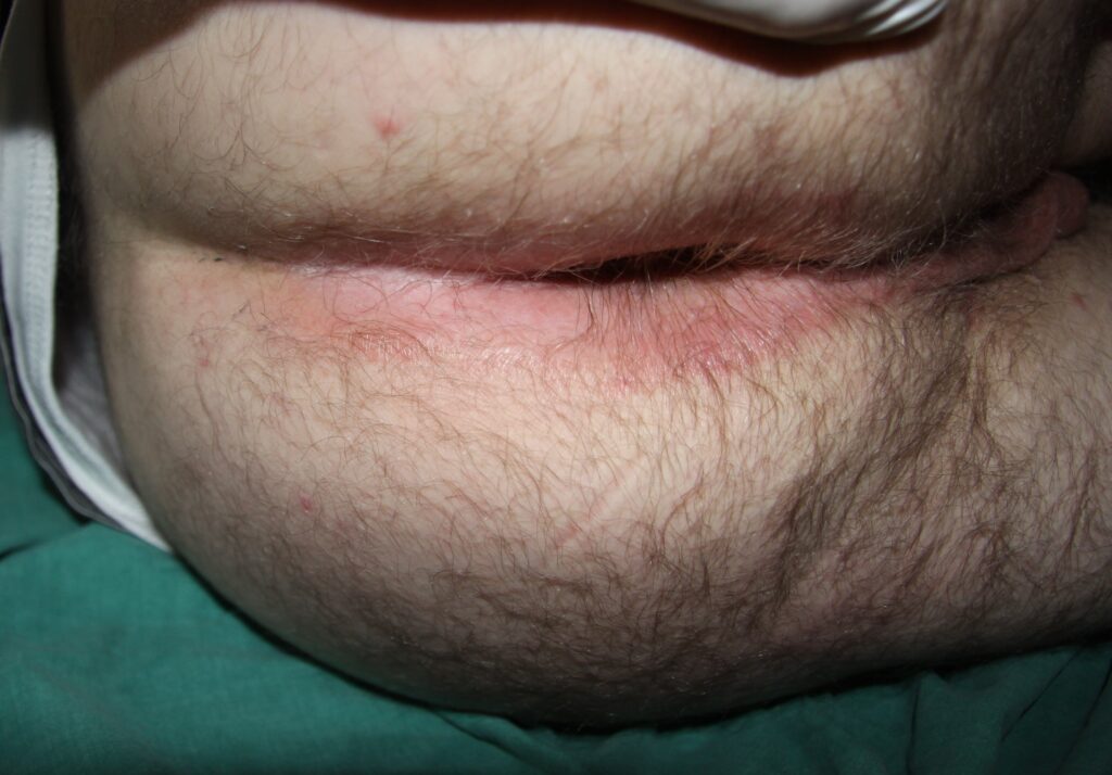
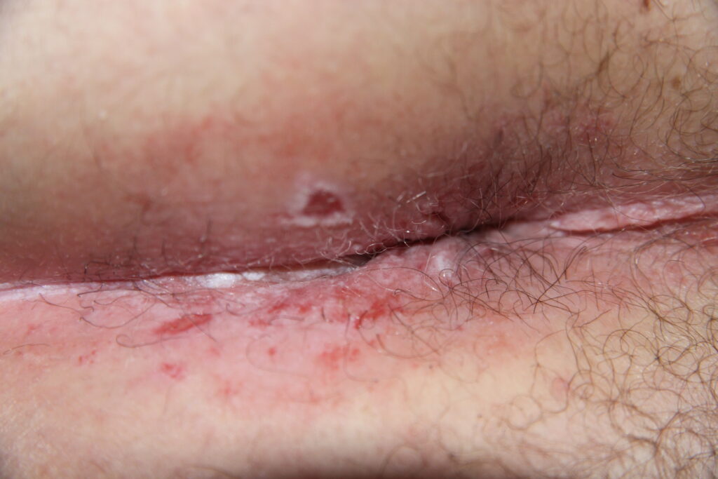
Irritant contact dermatitis is the most common cause of anogenital itch. The anogenital area is chronically exposed to many irritants. Repeated contact with skin irritants results in an innate immune (not allergic) response leading to inflammation (dermatitis). Irritants include anal secretions, faeces, urine, and sweat.
Attempts by the patient to relieve the itch often result in further irritation, especially from overzealous washing with soaps, shampoos, and wipes. Antifungal creams, topical antibacterials, and over-the-counter anti-itch creams may also irritate. Older patients with urinary incontinence are at particular risk.
The term incontinence-associated dermatitis (IAD) is increasingly promoted to highlight the vital need for good continence management when treating this problem. Older people’s skin is inherently vulnerable due to epidermal thinning, slow healing time, and a deficient skin barrier, all leading to increased risk of developing irritant contact dermatitis.
Anogenital pruritus is considered idiopathic when no dermatological cause is found. The pathogenesis is thought to be primarily due to faecal or urinary contact with the skin (both contain irritants and allergens). Repeated scratching and rubbing result in the clinical appearance of redness, excoriations, and lichenification. Moist skin folds are prone to the development of macerations and erosions.
Indeed, these findings are commonly found when the itch-scratch cycle is precipitated by an underlying dermatosis such as psoriasis or dermatitis (secondary pruritus). Added to all this, over washing and the application of over-the-counter treatments may result in contact irritant or allergic dermatitis. Making a diagnosis is not easy. A good strategy if you are not sure of an exact diagnosis is to initially manage the rash as an irritant contact dermatitis. In my experience, this approach will give a satisfactory outcome for the majority of patients presenting with anogenital pruritus.
Management of irritant contact dermatitis
Cleaning away faeces and urine is essential. It should be done gently, but thoroughly. The area should be dabbed (not rubbed) dry after washing. Soaps and shampoos are avoided for this purpose, but if used they must be thoroughly rinsed off before drying. A moisturising cream should be used as a soap substitute. After cleansing, an ointment moisturiser with barrier properties, such as Vaseline, should be applied. It might be applied to the perianal skin before the bowels are opened, before a shower, and before swimming. Loose cotton underwear is advised.
Our aim is to break the itch-scratch cycle. To settle the inflammation that is driving the pruritus, a topical steroid is best. Because of the risk of increased absorption by the anogenital skin, use the mild potency hydrocortisone ointment or the moderate potency clobetasone butyrate ointment. Compared with creams, ointments have less preservatives, lowering the risk of allergic contact dermatitis. Ointments also contribute a barrier effect.
However, if the patient prefers a cream, it is acceptable to prescribe one. In general, a topical steroid should not be continued indefinitely. If the problem recurs, a suppressive regime – clobetasone butyrate ointment applied on two days each week (such as Saturday and Sunday) – is thought to be safe in the medium- to long-term.
Although the risk of skin atrophy is high in the anogenital area, a short course of a potent topical steroid (betamethasone butyrate) may be used if inflammation is severe. Subsequently, the potency can be reduced to a moderate or mild potency topical steroid.
If treatment response is poor, consider possible explanations:
- Exposure to irritants has persisted. Failure to deal with incontinence when incontinence-associated dermatitis is the cause of the dermatitis is essential.
- An underlying dermatosis, eg, psoriasis, is present and needs specific treatment modalities such as a topical vitamin D analogue. The moist, occluded environment frequently alters the morphology of skin conditions, such as psoriasis, making the diagnosis difficult.
- Anogenital skin is prone to bacterial and fungal colonisation and infection. Dermatoses in this area are often secondarily infected. If the above measures are not working, consider the possibility of secondary infection. Hydrocortisone can be combined with antibacterials or antifungals, which carry a risk of allergic contact dermatitis and should be limited to seven days use before reverting to a steroid-only application.
POTENCY OF TOPICAL CORTICOSTEROID PREPARATIONS
Mild (Class I) – Hydrocortisone
Moderate (Class II) – Clobetasone butyrate
Potent (Class III) – Betamethasone butyrate
Supra Potent (Class IV) – Clobetasol propionate
(Class IV is 600 times more potent than Class I)
Allergic contact dermatitis
In contrast to irritant contact dermatitis, allergic contact dermatitis is much less common. It is caused by an allergic reaction to a substance that is in contact with the skin. Allergic contact dermatitis is a type IV (delayed) hypersensitivity reaction, which typically develops one to three days after exposure to an allergen. Sensitisation usually persists lifelong.
Clinical findings vary from redness, vesicles, oozing, and crusting (acute dermatitis) to scaling and lichenification (chronic dermatitis). On the anogenital skin, the commonest contact allergens include anaesthetics (benzocaine), fragrances (balsam of Peru), nickel, neomycin, fusidic acid, nystatin, latex (condoms), hydrocortisone, imidazoles (clotrimazole, ketoconazole), and methylisothiazolinone (now banned).
The diagnosis is definitively made by patch testing. Unfortunately, waiting times for testing are very long. Patients can also perform a repeated open application test by applying the suspected product to a small spot on the inner aspect of the upper arm, twice daily for several days, and then observing for a reaction.
Of course, a positive result only suggests the product is culpable. It does not identify what constituent of the product is the allergen. If the causative allergen is identified and is no longer applied to the skin, the condition is cured. When faced with a clinical picture of dermatitis and without access to patch testing, one might just ask the patient to stop all agents they have been applying and start a regime as outlined above for irritant contact dermatitis.
Allergy to a topical steroid (most commonly hydrocortisone) is a particular problem that is often not suspected because the patient gets relief once the steroid is applied. They may then continue applying or reapplying the cream resulting in persistence of the allergic contact dermatitis. The outlook is usually good as long as the patient adheres to allergen avoidance.
Psoriasis
Psoriasis involving the body flexures – such as the axillae, under female breasts, and behind the ears – significantly affect the clinical features of the disease compared with chronic plaque psoriasis on other body areas. These changes result from the effects of skin occlusion at these sites.
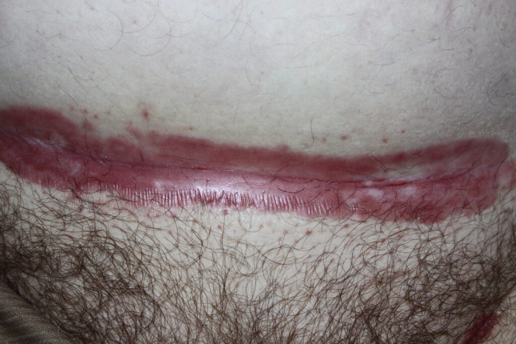
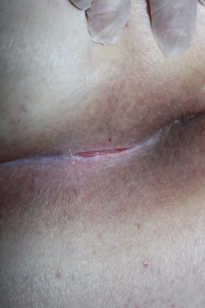
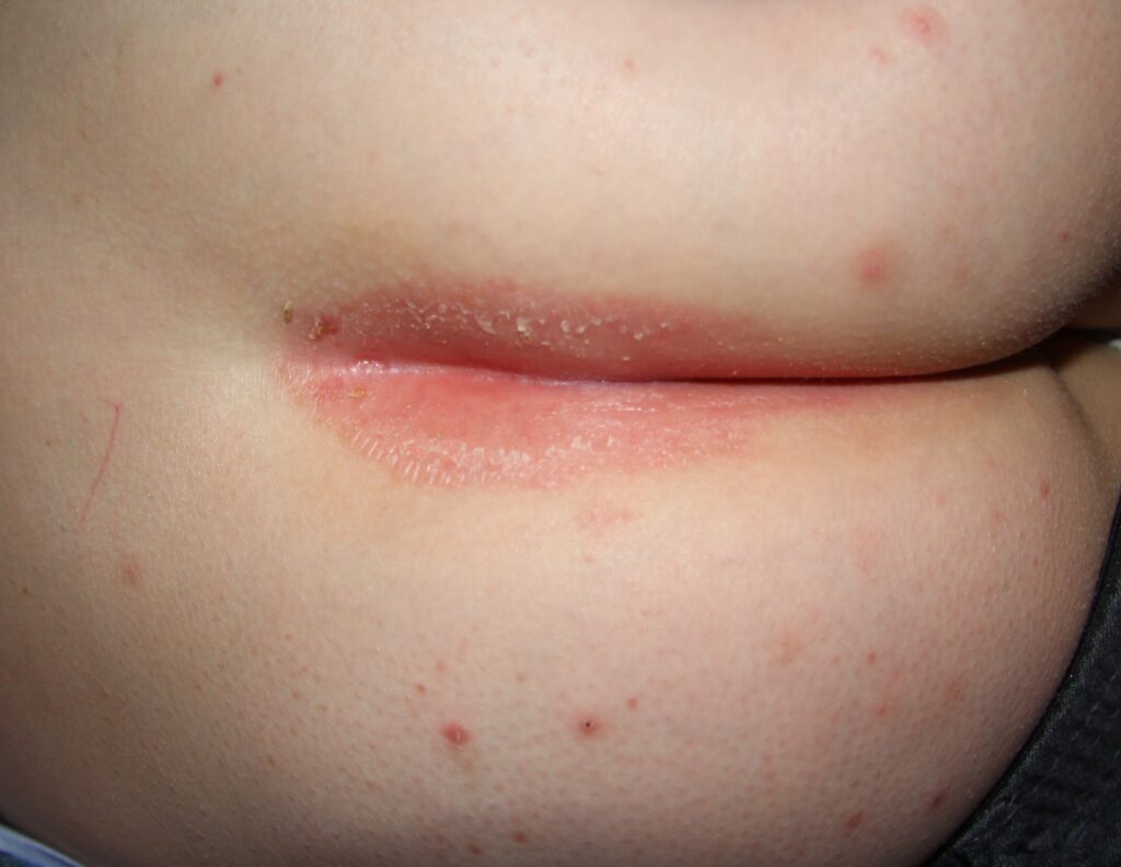
The most common regions for the development of intertriginous psoriasis (also called inverse pattern psoriasis or flexural psoriasis) are in the inguinal folds and natal clefts (also called gluteal cleft) of the anogenital region. Occlusion results in marked changes in the clinical picture. Examination reveals sharply demarcated, but thin, plaques that are pink to red in colour, have no scales or just a few macerated ones, a shiny, glaze-like surface, and a central fissure at the apex of the skin fold.
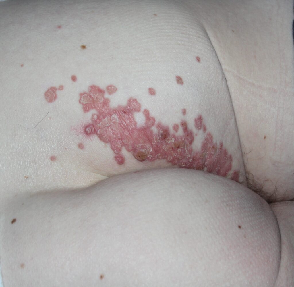
In contrast, non-occluded plaques of the skin outside the flexures have the features of chronic plaque psoriasis, being well demarcated, thickened, pink in colour, and covered in silvery scales.
Psoriasis may be itchy, and repeated rubbing and scratching may lead to a lichenified eczema that will, in turn, itch even more – driving the itch-scratch cycle. Sexual difficulties, embarrassment, and psychological distress are common. Furthermore, flexures are more prone to infections such as dermatophytes, candida, and bacteria which can trigger a psoriasis flare.
When treating anogenital psoriasis, effective topical preparations are limited. Best results are achieved by combining an emollient regime with the moderately potent topical steroid clobetasone ointment, applied in the morning with the topical vitamin D analogue, calcipotriol ointment, applied at night.
Potent and very potent topical steroids should be avoided on all anogenital skin, whether flexural or non-flexural, due to the risk of skin atrophy. If an acceptable result is achieved by four weeks, the frequency of clobetasone application can be reduced to twice weekly for a couple of weeks and then stopped.
Prolonged use of any potency topical steroid risks the development of tachyphylaxis, ie, lessening of the therapeutic response with constant use. Calcipotriol ointment frequently irritates. The anti-inflammatory effect of a topical steroid ameliorates this irritation. Calcipotriol can be continued at a frequency of application that controls the disease. Tacrolimus has been used as an alternative with good results over many years. However, this is an off-label use of tacrolimus.
These treatment regimens are not very effective on the thick plaques of psoriasis on other body sites. Occlusion at flexural sites leads to increased absorption and potency of topical applications, resulting in a better therapeutic response with weaker potency applications.
Seborrhoeic dermatitis
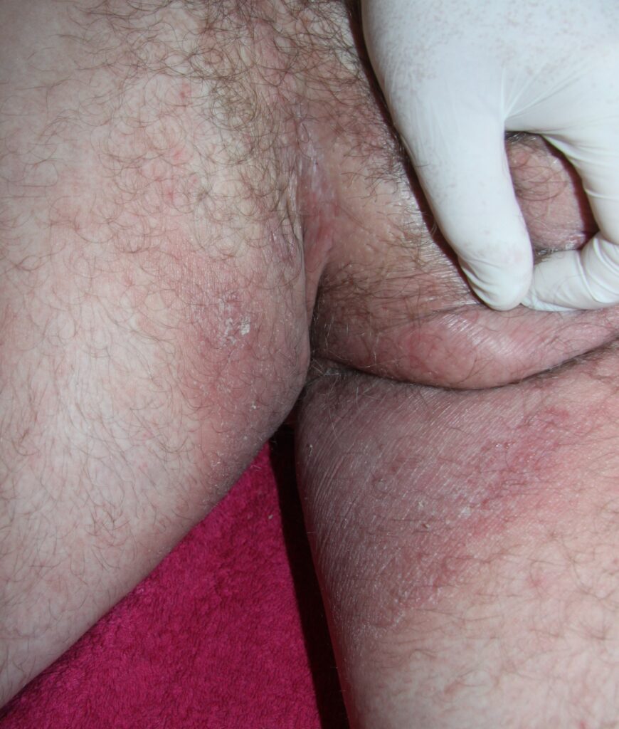
Seborrhoeic dermatitis is very common, running a chronic, mild course. It presents on areas with high sebum production and the body folds. Its pathogenesis is not fully defined. It may be related to seborrhoea and the yeast Malassezia furfur (Pityrosporum ovale). The adult form of seborrhoeic dermatitis develops after puberty when the sebaceous glands become active. In the anogenital region, well demarcated, red-brown patches develop.
Pruritus may be mild or moderate. Outside the flexures, bran-like scale may be found. To help with confirming the diagnosis, check the scalp, the nasolabial folds, eyes, ears, the pre-sternal area, and other body flexures. Clinical appearance may overlap with psoriasis, a condition known as sebopsoriasis.
As in psoriasis, fissuring at the apex of the skin fold can occur. The moist environment of the skin folds and the exposure to irritants frequently lead to a combination of irritant contact dermatitis and seborrhoeic dermatitis. The lesions appear more a bright red colour and erosions are frequent.
Irritant avoidance and emollient use, as outlined above, are recommended for all cases. Topical azoles (clotrimazole, miconazole, and ketoconazole) have a high efficacy. A maintenance regimen of a topical azole may be needed as relapse is common. Hydrocortisone, added to miconazole or clotrimazole in a fixed dose combination product (Daktacort, Canesten HC), is indicated if inflammation is severe.
Atopic eczema
The primary features of atopic eczema are a poor skin barrier and itch. It is not surprising therefore that atopic patients frequently present with pruritus and eczematous rash in the anogenital area. Atopic patients are particularly susceptible to irritant exposure and will frequently present with irritant contact dermatitis superimposed on atopic dermatitis.
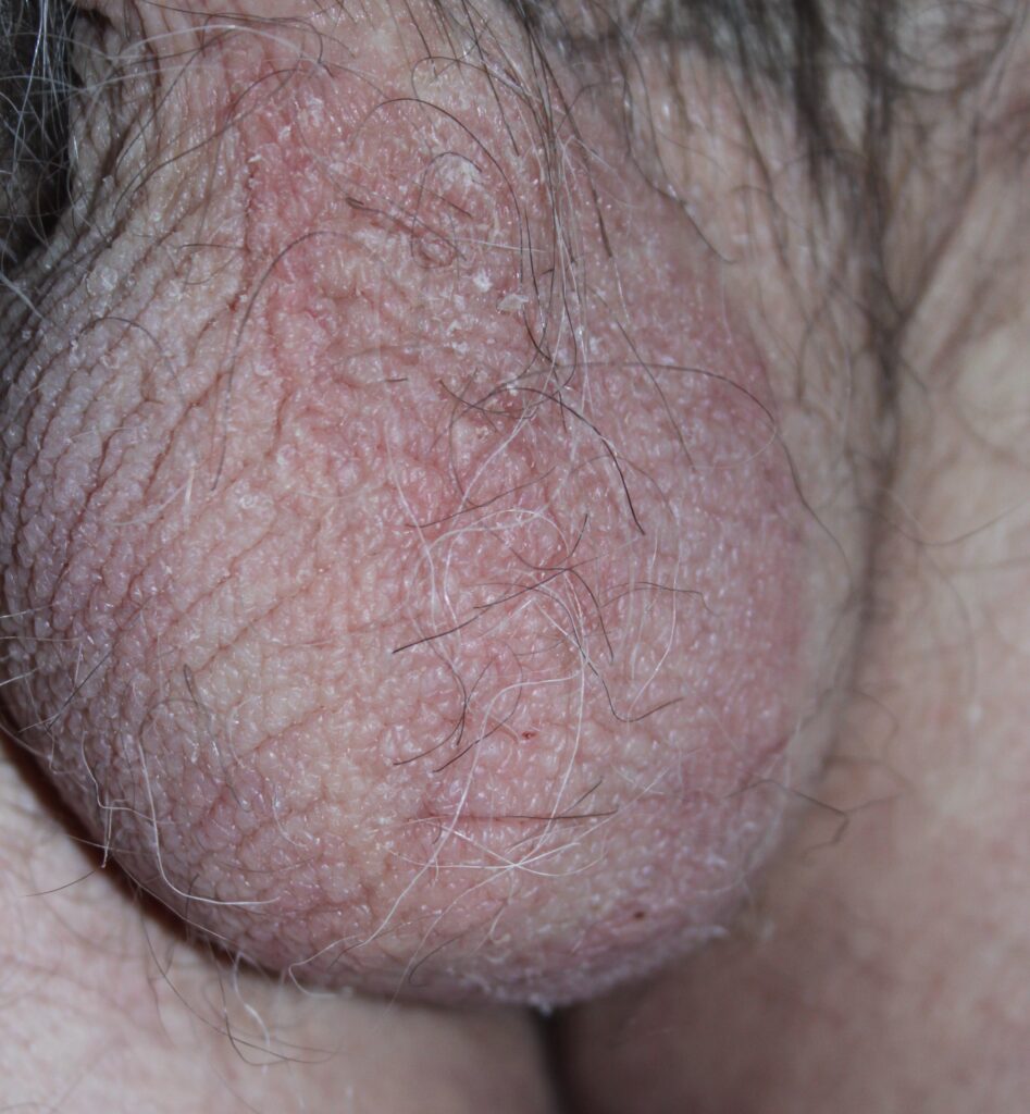
Severe pruritus involving the genital skin sets off an itch-scratch cycle, resulting in lichenified skin. Scrotal skin is particularly susceptible. Management is essentially the same as for irritant contact dermatitis, with a particular emphasis on irritant avoidance and moisturisation.
Candidiasis
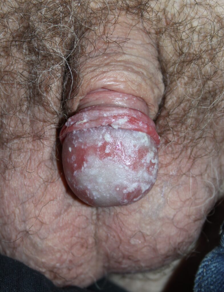
Cutaneous candida infection is usually a secondary opportunistic phenomenon rather than the primary cause of a dermatosis. Contact dermatitis, psoriasis, and seborrhoeic dermatitis may be involved. Affected areas may have a curdy, whiteish discharge – ‘cottage cheese like’ – due to maceration of scales. Patients with diabetes who are on a broad-spectrum antibiotic or oral steroid therapy are at increased risk.
Topical azoles may be effective. Fluconazole, given as a stat 150mg dose may be needed for more severe disease. Candida persistence and recurrence is common. Some advocate Fluconazole as 150mg at 72-hour intervals if the stat dose is ineffective.
-
- Figure 1
-
- Figure 2
-
- Figure 3
-
- Figure 4
-
- Figure 5
-
- Figure 6
-
- Figure 7
-
- Figure 8
-
- Figure 9
-
- Figure 10
-
- Figure 11
-
- Figure 12
-
- Figure 13
FIGURE 1: When faced with a pruritic rash in the anogenital flexures, it is difficult to determine exactly what the diagnosis is. Often, we get irritant or allergic contact dermatitis superimposed on seborrhoeic dermatitis, psoriasis or atopy. This patient was not applying anything to the rash and skin examination revealed no sign of any other dermatosis. A diagnosis of irritant contact dermatitis was made.
FIGURE 2: A more severe form of irritant contact dermatitis with erosions and oozing.
FIGURE 3: This patient complains of severe itch. Examination shows marked lichenification, redness, and slight scaling. This is lichen simplex chronicus. Most of these patients have probably got underlying atopy.
FIGURE 4: Like all dermatitis in the anogenital area, it can be difficult to put an exact label on this pruritic, red, and slightly scaly rash. General skin examination supported a diagnosis of seborrhoeic dermatitis.
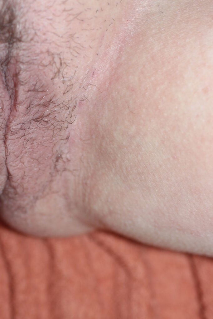
FIGURE 5: A similar situation as in Figure 4, but in a female patient. Seborrhoeic dermatitis.
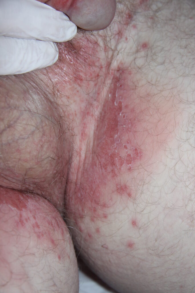
FIGURE 6: Overvigorous washing with soaps and shampoos have led to the development of irritant contact dermatitis.
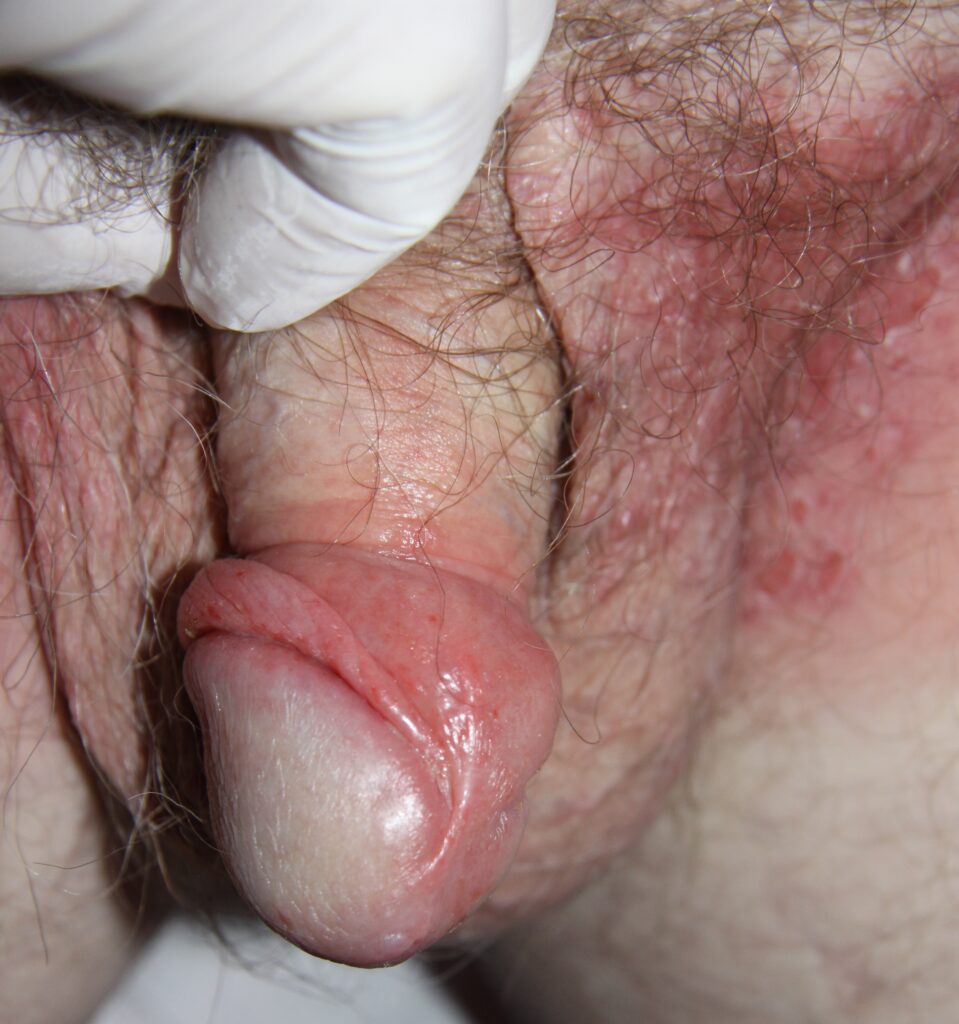
FIGURE 7: Allergic contact dermatitis to fucidic acid. The cream was applied to the genital skin, but some got on the inner, upper thigh. Fucidic acid commonly sensitises.
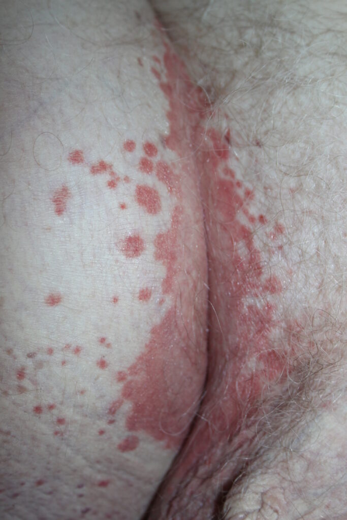
FIGURE 8: This patient had signs on the knees and elbows confirming the diagnosis of psoriasis. The satellite plaques are thicker and have some scale, in contrast to the thin, glazed plaques in the skin fold. The satellite plaques need to be differentiated from the satellite lesions in candida infection.
FIGURE 9: Flexural psoriasis of a lower abdominal skin fold, just above the groin. Note the discrete border, thin plaque, absence of scale, glazed surface, and early fissuring at the apex of the skin fold.
FIGURE 10: This has psoriasis of the gluteal cleft, which is healing with post-inflammatory hyperpigmentation. Fissuring at the apex of the skin fold is seen.
FIGURE 11: Plaques of psoriasis on the buttock. In contrast to psoriasis in the skin folds the plaques, while still discrete, are thicker and have scale, which is better demonstrated by gently scratching the surface.
FIGURE 12: A teenage patient with psoriasis of the gluteal cleft. Note the macerated scales at the outer edge of the plaques.
FIGURE 13: Candida infection of the glans penis. Note the thick white exudate, yellowish pustules and erosions. The exudate in places forms a pseudo membrane over the glans.