Reference: June 2026 | Issue 6 | Vol 12 | Page 37
Osteoporosis is a preventable skeletal disease, characterised by reduced bone strength and an increased susceptibility to fragility fractures. Osteoporosis is often a silent disease until the first fragility fracture. But the good news is that effective strategies exist to identify and treat osteoporosis early, reducing the risk of fractures and related complications.
This article provides an overview of osteoporosis risk factors, screening, relevant investigations and management, based on the latest guidelines and practical clinical considerations.
Epidemiology
Osteoporosis represents a major public health concern and despite advances in diagnosis and treatment, it remains underdiagnosed and undertreated. Fragility fractures are associated with chronic pain and disability, loss of independence, increased mortality, and substantial healthcare costs.
Approximately 50 per cent of women and 20 per cent of men over the age of 50 will sustain a fragility fracture during their lifetime. In the UK, over 500,000 fragility fractures occur each year, while an estimated two million fractures occur in the US per annum. Due to the aging population, these staggering numbers are expected to rise significantly, with hip fracture incidence projected to increase by 20 per cent by 2030 and by 50 per cent by 2050.1,2,3
Screening
Who to screen
Screening strategies aim to identify individuals at increased fracture risk before clinical events occur. Most international guidelines, including those from the National Osteoporosis Guideline Group and National Osteoporosis Foundation, recommend screening all women aged ≥65 years, postmenopausal women, and men aged ≥50 with risk factors, individuals with prior fragility fractures, and those on long-term glucocorticoids. Routine screening is generally not recommended in adults <50 without significant risk factors. There is less consensus for universal screening in men aged ≥70, although clinical judgment is advised.4,5
How to screen
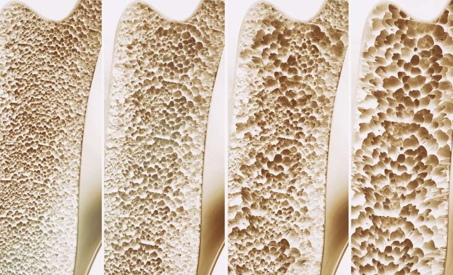
The gold standard diagnostic screening tool for evaluating bone mineral density (BMD), which is used as a proxy to bone strength, is dual-energy x-ray absorptiometry (DXA). BMD is reported as a comparison score (T-score), where the patient’s BMD is compared to a healthy 20- to 30-year-old reference population of the same sex. T-score is expressed in standard deviation (SD) from the ideal peak bone mass, with a T-score of greater than -1 classified as normal, between -1 and -2.5 as osteopenia, and greater than -2.5 as osteoporosis.
A DXA scan is an excellent predictor of fracture. For every SD decrease in age adjusted BMD, there is a two-fold increase in fracture risk. Despite this however, a clear distinction between osteopenia and osteoporosis is considered arbitrary and those with advanced osteopenia face similar risks of fragility fractures as others with osteoporosis by T-score definition. This highlights the limitations of using a T-score alone in risk stratifications and the need to incorporate additional risk factors independent of T-score to stratify patients’ fracture risk.
One of the most commonly used fracture risk prediction tools is the fracture risk assessment tool (FRAX). FRAX incorporates 12 fracture risk factors to estimate a 10-year probability of hip and major osteoporotic fracture. Such tools help guide treatment decisions, particularly in patients with osteopenia.4
Risk factors
Osteoporosis is multifactorial. The following list highlights some of the many risk factors.6
Clinical factors
▶ Age
▶ Previous fragility fracture
▶ Family history of osteoporosis
▶ Low body mass index
▶ Height loss or kyphosis.
Lifestyle factors
▶ Smoking
▶ Excess alcohol intake
▶ Low calcium intake
▶ Sedentary lifestyle.
Medications
▶ Glucocorticoids
▶ Aromatase inhibitors
▶ Anti-convulsant.
Secondary causes
▶ Inflammatory diseases (eg, rheumatoid arthritis, inflammatory bowel disease)
▶ Endocrine disorders (eg, hypogonadism, hyperthyroidism)
▶ Chronic kidney disease.
Investigations
Once osteoporosis or a fragility fracture is identified, the next step is to look for secondary contributors. This is especially important in younger patients, men, those with unexpectedly severe osteoporosis, or when the pattern of fracture seems disproportionate to age and risk factors.
Baseline routine tests typically include full blood count, renal profile, calcium, phosphate, alkaline phosphatase, 25-hydroxy vitamin D, and parathyroid hormone, with further targeted tests such as thyroid function, sex hormone profile, prolactin, coeliac serology, and serum protein electrophoresis.4
Management
Non-pharmacological measures
Management always begins with lifestyle and non-pharmacological measures. These should be recommended to all patients, regardless of whether they also need drug therapy. Adequate calcium intake is important for bone health, and a total intake of about 1,000-1,500mg daily from diet and supplements combined is generally advised. Vitamin D should also be optimised, with supplementation of around 800IU daily commonly used to support calcium absorption, maintain bone mineralisation, and reduce the risk of deficiency. These recommendations are often individualised depending on age, dietary intake, and individual risk factors.
Regular weight-bearing and resistance exercise should be encouraged because it helps maintain bone strength, improves muscle function, and supports balance and mobility. Smoking cessation is important, as smoking is associated with lower bone density and higher fracture risk. Alcohol intake should be moderated, since excess alcohol can adversely affect bone health and increase falls risk. In addition, falls prevention strategies are a key part of osteoporosis care and may include review of vision, footwear, home hazards, gait and balance, sedating medications, and the use of aids where appropriate.4,5,6
Pharmacologic treatment
Despite optimising non-pharmacologic measures, a great number of patients require pharmacologic treatment. Pharmacologic treatment is indicated in patients with fragility fractures, those with osteoporosis on DXA defined by a T-score of less than or equal to -2.5, and in high-risk individuals identified using FRAX assessment. Additionally, there is particular emphasis on early treatment in glucocorticoid-induced osteoporosis, given the rapid bone loss that can occur with steroid exposure.
Anti-resorptive therapies:
Bisphosphonates remain the first-line therapy for most patients. They are well established, effective, and well tolerated. Additionally, availability in both oral and intravenous infusion formulations provides added flexibility in route of administration, which can be an important consideration in cases such as underlying oesophageal disease and poor oral tolerance.
Before starting bisphosphate therapy, vitamin D deficiency should be corrected and renal function should be assessed, as bisphosphonates should be avoided in severe renal impairment, typically when eGFR is below 30-35mL/min.
Denosumab is another important anti-resorptive. It is a targeted monoclonal antibody that prevents fractures by inhibiting bone breakdown. It is administered subcutaneously every six months. Unlike bisphosphonates, it is not renally excreted, which makes it particularly useful in patients with renal impairment or intolerance to oral bisphosphonates.
Selective oestrogen receptor modulators (such as raloxifene) and hormone replacement therapy (HRT) also have anti-resorptive effects on the bone and have a role in carefully selected patients.
Anabolic therapies:
Anabolic agents improve BMD by stimulating bone formation. These agents are usually reserved for patients with very high fracture risk (T-score ≤−3.5), those with multiple fractures, or failure of previous anti-resorptive therapy. Anabolic agents are highly effective in appropriately selected patients; however, they are approved for use only for a limited period and should be followed by an anti-resorptive agent to maintain the benefits once the anabolic course is completed.4,5,6
Monitoring
Routine monitoring of patients on bone therapies is an essential component of safe and effective management of osteoporosis. Treatment response should be evaluated regularly with repeat DXA scan every two to three years (depending on baseline fracture risk), the treatment being used, and whether the result is likely to influence management. In addition to imaging, follow-up should include assessment of adherence, tolerability, interval falls, and the occurrence of any new fragility fractures.
Treatment review is also the point at which clinicians should reconsider whether the current strategy remains appropriate. In patients receiving long-term bisphosphonate therapy, drug holidays may be considered for individuals with appropriate response and lower fracture risk.
Safety considerations
While serious adverse effects are rare, anti-resorptive agents, particularly bisphosphonates, can cause rare complications that need to be considered and thoroughly discussed with patients prior to and during the treatment course.
Osteonecrosis of the jaw and atypical femoral fractures are both uncommon, but are clinically important and warrant awareness. Underlying poor oral health, invasive dental procedures, smoking, diabetes, glucocorticoid use, and prolonged anti-resorptive exposure, increase the risk of osteonecrosis of the jaw. For that reason, preventive dental assessment and optimisation of oral health are recommended before treatment is started.
Atypical femoral fractures are also rare, but they are clinically significant as they may occur with prolonged anti-resorptive exposure and can present insidiously. Risk factors associated with atypical femoral fracture are dose and duration of exposure to anti-resorbtives, glucocorticoid use, Asian ethnicity, and co-morbid conditions such as diabetes.7,8,9
Denosumab has some additional safety considerations that differ from those of bisphosphonates. Hypocalcaemia may occur, especially in patients with vitamin D deficiency or impaired renal function, so calcium and vitamin D status should be optimised before treatment. Another key issue is the rapid bone loss that can follow cessation of denosumab, with increased risk of rebound vertebral fractures. Therefore transition to another anti-resorptive agent should be planned rather than stopping treatment abruptly.
Anabolic agents, such as teriparatide and abaloparatide, may cause hypercalcaemia and should be used with caution in patients with hyperparathyroidism or a history of renal stones. These agents are also contraindicated in patients with a history of skeletal radiation and bone cancers. The newest anabolic agent, romosozumab, carries an increased risk of myocardial infarction and stroke and should not be initiated in patients who have experienced a major adverse cardiovascular event within the preceding year.
Conclusion
Osteoporosis remains a significant and growing clinical challenge, particularly as populations age and the burden of fragility fracture continues to rise. Although effective therapies are available, a substantial treatment gap persists, especially in secondary prevention after fragility fracture, where many high-risk patients still go untreated.
Early identification, careful investigation for secondary causes, and timely initiation of appropriate therapy are essential if fracture risk is to be reduced meaningfully. Clinicians should take a proactive and guideline-based approach, while also tailoring management to the individual patient’s fracture risk, comorbidities, treatment preferences, and long-term safety considerations.
References
- Burge R, Dawson-Hughes B, Solomon DH, Wong JB, King A, Tosteson A. Incidence and economic burden of osteoporosis-related fractures in the United States, 2005–2025. J Bone Miner Res. 2007.
- Singh JA, et al. Data cited in your presentation regarding post-fracture treatment gap and imminent fracture risk. J Bone Miner Res. 2023.
- National Committee for Quality Assurance. The State of Healthcare Quality 2022.
- National Osteoporosis Guideline Group (NOGG). Clinical guideline for the prevention and treatment of osteoporosis. Updated 2024.
- American College of Rheumatology guideline materials on glucocorticoid-induced osteoporosis, as reflected in contemporary guideline summaries.
- LeBoff MS, Greenspan SL, Insogna KL, et al. The clinician’s guide to prevention and treatment of osteoporosis. Osteoporosis Int. 2022;33(10):2049-2102.
- Khan AA, Morrison A, Hanley DA, et al. Diagnosis and management of osteonecrosis of the jaw: A systematic review and international consensus. J Bone Miner Res. 2015.
- Shane E, Burr D, Abrahamsen B, et al. Atypical subtrochanteric and diaphyseal femoral fractures: Second report of a taskforce of the American Society for Bone and Mineral Research. J Bone Miner Res. 2014.
- Adler RA, El-Hajj Fuleihan G, Bauer DC, et al. Managing osteoporosis in patients on long-term bisphosphonate treatment: Report of a taskforce of the American Society for Bone and Mineral Research. J Bone Miner Res. 2016.