


Minimally-invasive procedure relieves tremors in Parkinson’s patients
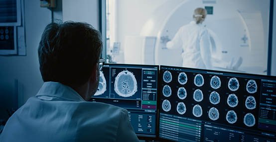
A procedure that applies pulses of focused ultrasound to the brain is safe and effective for reducing tremors and improving quality of life in people with essential tremor (ET) or Parkinson’s disease (PD) tremor, according to a new study presented at the 2019 Annual Meeting of the Radiological Society of North America (RSNA).
Established treatment options for reducing tremors in patients who have not responded to medical therapy include deep-brain stimulation, but a more recently available option is magnetic resonance-guided focused ultrasound (MRgFUS) thalamotomy, an incisionless interventional radiology procedure in which focused beams of sound energy are used to heat and destroy a small part of the thalamus. The procedure gives relief to the opposite side of the body.
As a minimally-invasive approach, focused ultrasound has advantages over deep-brain stimulation, including a reduced risk of complications from bleeding and infections, according to the study lead author Dr Federico Bruno, a radiologist in the Department of Biotechnological and Applied Clinical Sciences at the University of L’Aquila in L’Aquila, Italy.
“Another advantage is the immediate effect this treatment provides, unlike deep-brain stimulation, which requires a break-in period for the electrostimulation,” he said. “Additionally, treatment with MRgFUS requires shorter hospitalisation and is a fairly well-tolerated procedure, even by more fragile patients.”
For the new study, Dr Bruno and colleagues enrolled 39 patients, average age 64.5 years, with disabling tremors that had not responded to treatment. The people in the study group, including 18 with ET and 21 with PD, had experienced symptoms for an average of more than 10 years.
They found that 37 of 39 patients, or 95 per cent, had substantial and immediate reduction of tremor. These reductions in tremor were sustained in follow-up evaluations. Quality-of-life evaluation showed substantial improvement in both the ET and PD groups.
“The study we presented reports our experience of over a year in the treatment of tremor by thalamotomy with focused ultrasound,” Dr Bruno said. “It is worth noting that we had a high number of patients with Parkinson’s disease in our series, compared to previously published data, where the procedure was used mainly in the treatment of essential tremor patients.”
Currently, MRgFUS thalamotomy is only available at a limited number of sites worldwide, Dr Bruno said, but may become more widespread as research findings supporting its use are published. Improvements in neuroimaging techniques that allow for greater precision and detail in planning, implementation and monitoring over time of the treatment should also expand its availability.
Future research in this area includes the possibility of treating both sides of the thalamus. MRgFUS is also being explored in areas beyond movement disorders, Dr Bruno said. Several preclinical studies and clinical trials are looking at the technique for the treatment of other neurological conditions like neuropathic pain, epilepsy and obsessive-compulsive disorders, as well as for treatment of brain tumours.
Novel MRI-guided ultrasound treatment for prostate cancer
A novel MRI-guided procedure that uses therapeutic ultrasound effectively treats prostate cancer with minimal side-effects, according to a new study presented at the 2019 Annual Meeting of the RSNA.
Researchers said the incision-free technique, MRI-guided transurethral ultrasound ablation (TULSA), could also be used to treat benign enlargement of the prostate gland.
TULSA delivers precise doses of sound waves to diseased prostate tissue while sparing the healthy nerve tissue surrounding the prostate.
The novel rod-shaped device that is inserted into the urethra has 10 ultrasound-generating elements that can cover the entire prostate gland.
The elements are controlled automatically by a software algorithm that can adjust the shape, direction and strength of the therapeutic ultrasound beam and the entire procedure takes place in an MRI scanner.
“Unlike with other ultrasound systems on the market, you can monitor the ultrasound ablation process in real time and get immediate MRI feedback on the thermal dose and efficacy,” explained study co-author Prof Steven S Raman, Professor of Radiology and Urology, and Director of Prostate MR Imaging and Interventions and Prostate MR Imaging Research at the University of California at Los Angeles (UCLA). “It’s an outpatient procedure with minimal recovery time.”
In the new multi-centre study, researchers reported on the 12-month outcomes from the TULSA-PRO ablation clinical trial (TACT). The trial enrolled 115 men, median age 65 years, with localised low- or intermediate-risk, gland-confined prostate cancer. Clinicians delivered TULSA treatment to the entire gland.
Prostate volume in the study group decreased on average from 39 cubic centimetres pre-treatment to 3.8 cubic centimetres one year after treatment. Overall, clinically significant cancer was eliminated in 80 per cent of the study participants. Out of 111 men, 72 (65 per cent) had no evidence of any cancer at biopsy after one year. Blood levels of PSA fell by a median of 95 per cent. There were low rates of severe toxicity and no bowel complications.
“We saw very good results in the patients, with a dramatic reduction of over 90 per cent in prostate volume and low rates of impotence, with almost no incontinence,” Dr Raman said.
Approved for clinical use in Europe, TULSA has just received FDA 510(k) clearance for prostate tissue ablation in the US. Assuming follow-up studies support the preliminary results, the technique could develop into an important tool for treating both prostate cancer and benign prostatic hyperplasia, or enlargement of the prostate.
“There are two very unique things about this system,” Dr Raman said. “First, you can control with much more finesse where you’re going to treat, preserving continence and sexual function. Second, you can do this for both diffuse and localised prostate cancer and benign diseases, including benign hyperplasia.”
TULSA also has the benefit of allowing further treatment if needed, Dr Raman said.
The study also supports the use of MRI for post-treatment monitoring of patients who undergo TULSA.
MRI at one year after treatment had a negative predictive value of 93-to-96 per cent for detecting residual cancer, meaning it was very accurate for ruling out disease recurrence in patients.
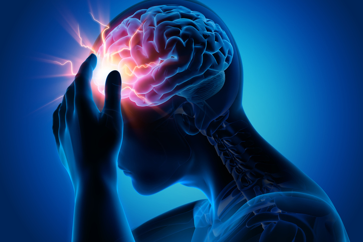
Focused ultrasound may open door to Alzheimer’s treatment
Focused ultrasound is a safe and effective way to target and open areas of the blood-brain barrier, potentially allowing for new treatment approaches to Alzheimer’s disease, according to initial study results presented at the 2019 Annual Meeting of the RSNA.
There currently is no effective treatment for Alzheimer’s disease, the most common cause of dementia. Studies on animals have shown that pulses of low-intensity focused ultrasound (LIFU) delivered under MRI guidance can reversibly open this barrier and allow for targeted drug and stem-cell delivery.
Researchers at three sites have been studying LIFU in humans for more than a year in a clinical trial led by Dr Ali Rezai, Director of the West Virginia University (WVU) Rockefeller Neuroscience Institute. For the new study, researchers delivered LIFU to specific sites in the brain critical to memory in three women, ages 61, 72 and 73 years, with early-stage Alzheimer’s disease and evidence of amyloid plaques (abnormal clumps of protein in the brain that are linked with Alzheimer’s disease). The patients received three successive treatments at two-week intervals. Researchers tracked them for bleeding, infection and oedema, or fluid build-up.
“The results are promising,” said study co-author Dr Rashi Mehta, Associate Professor at WVU and research scholar at West Virginia Clinical and Translational Science Institute. “We were able to open the blood-brain barrier in a very precise manner and document closure of the barrier within 24 hours. The technique was reproduced successfully in the patients, with no adverse effects.”
MRI-guided LIFU involves placement of a helmet over the patient’s head after they are positioned in the MRI scanner. The helmet is equipped with more than 1,000 separate ultrasound transducers angled in different orientations. Each transducer delivers sound waves targeted to a specific area of the brain. Patients also receive an injection of a contrast agent made up of microscopic bubbles. Once ultrasound is applied to the target area, the bubbles oscillate, or change size and shape.
“The helmet transducer delivers focal energy to specified locations in the brain,” Dr Mehta explained. “Oscillation of the microbubbles causes mechanical effects on the capillaries in the target area, resulting in a transient loosening of the blood-brain barrier.”
LIFU could help deliver therapeutic drugs into the brain to improve their effectiveness. Even without drugs, opening of the brain-blood barrier in animals has shown positive effects, Dr Mehta said.
While the research so far has focused on the technique’s safety, in the future the researchers intend to study LIFU’s therapeutic effects.
“We’d like to treat more patients and study the long-term effects to see if there are improvements in memory and symptoms associated with Alzheimer’s disease,” Dr Mehta said. “As safety is further clarified, the next step would be to use this approach to help deliver clinical drugs.”
Imaging reveals the brain pathways behind depression
MRI illuminates abnormalities in the brains of people with depression, potentially opening the door to new and improved treatments for the disorder, according to two studies presented at the annual meeting of the RSNA.
Major depressive disorder (MDD) is one of the most common and debilitating mental disorders worldwide and limited understanding of the brain changes associated with MDD hinders the effectiveness of treatments.
“Unfortunately, with current treatments there is a large chance of relapse or recurrence,” said Dr Kenneth T Wengler, PhD, from Columbia University in New York City and co-author of one of the studies. “To develop new, more effective treatments, we must improve our understanding of the disorder.”
Dr Wengler and colleagues recently studied connections between MDD and disruptions in the blood-brain barrier (BBB). Using a new MRI technique, they developed intrinsic diffusivity encoding of arterial labelled spins (IDEALS), looked at BBB water permeability, or the movement of water out of the blood vessels and into the brain tissue.
Comparison of results in 14 healthy individuals and 14 MDD patients found that less water moved from inside the blood vessels to outside in the MDD patients, representing disrupted BBB integrity. This difference was particularly large in two regions of the brain: The amygdala and the hippocampus.
“We observed disruption of the blood-brain barrier in grey matter regions known to be altered in major depressive disorder,” Dr Wengler said. “This study helps improve our understanding of the pathophysiology of depression and can open new avenues of treatment for a disorder that affects over 100 million individuals worldwide.”
A second study presented at RSNA 2019 looked at abnormalities in the complex network of connections in the brain known as the connectome for their role in depression. Previous research has focused on characterising the connections between different brain regions, but this study, from researchers at the University of North Carolina (UNC), looked deeper within individual brain regions.
The researchers compared 66 adults with MDD and 66 matched healthy controls during wakeful rest using functional MRI (fMRI) and a newly-developed multi-scale neural model inversion framework that linked the brain’s microscopic circuitry with its larger-scale interactions. As part of the study, the researchers were able to assess excitatory or inhibitory influence between neuronal cell groups. A proper balance between excitation and inhibition is crucial for a well-functioning brain.
Patients with MDD had abnormal patterns of excitation and inhibition at the dorsal lateral prefrontal cortex, a brain area important to cognitive control functions, including the regulation of the amygdala, a key region embedded deep in the brain for expression of emotion. It has long been hypothesised that malfunctioning inhibitory control over the amygdala could result in depressive symptoms.
“In our study, we found that excitation and inhibition in the brain regions in control of executive functions and emotional regulation were reduced in patients with MDD,” said study co-author Dr Guoshi Li, PhD, from the Image Display, Enhancement and Analysis (IDEA) group at UNC. “This suggests that control functions in MDD are impaired, which may lead to elevated responses in the amygdala, resulting in increased anxiety and other negative moods.”
In addition, the researchers found that recurrent excitation in the thalamus, an area of the central brain that is also responsible for emotional regulation, was abnormally elevated in patients with MDD.
Dr Li said the new approach could open the door for a deeper understanding of the mechanisms behind depression.
“Current methods of studying the brain provide a superficial understanding of connectivity,” Dr Li said. “This method allows us to identify impaired connectivity within each brain region, making it a potentially more powerful tool to study the neuromechanism of brain disorders and develop more effective diagnosis and treatment.”
AI helps find signs of heart disease on lung cancer CT screens
Artificial intelligence (AI) provides an automated and accurate tool to measure a common marker of heart disease in patients getting chest CT scans for lung cancer screening, according to a study presented at the 2019 Annual Meeting of the RSNA.
While low-dose chest CT scans are intended to diagnose lung cancer, coronary artery calcium is also visible on CT and cholesterol guidelines encourage using the calcium score to help physicians and patients decide whether to take a statin, noted study co-senior author Dr Michael T Lu, Director of AI in the Cardiovascular Imaging Research Centre (CIRC) at Massachusetts General Hospital (MGH) in Boston.
However, despite its prognostic value, coronary artery calcium is not routinely measured in low-dose CT lung screening, as the measurements require dedicated software and add time to the interpretation.
“If our tool detects a lot of coronary artery calcium in a patient, then maybe we can send that patient to a specialist for follow-up,” said lead author Dr Roman Zeleznik, MSc, BSc, from the Artificial Intelligence in Medicine (AIM) Programme at Boston’s Brigham and Women’s Hospital (BWH) and Dana-Farber Cancer Institute. “This would make it easier for patients to get appropriate treatment.”
The research team, which represents a close collaboration between MGH’s CIRC and AIM at BWH, recently developed and tested a technique that uses deep learning to automatically measure coronary artery calcium on chest CT images. They trained the deep learning system on cardiac CTs and chest CTs in which the coronary artery calcium had been measured manually.
They then tested the system on CT scans from thousands of heavy smokers, aged 55-to-74 years, who were part of the US National Lung Screening Trial (NLST), a major study that established CT’s value in providing early detection of lung cancer.
The results showed that the deep learning-derived coronary artery calcium scores corresponded closely to those of human readers. In addition, there was a significant association between deep learning calcium scores and cardiovascular death over follow-up of 6.5 years.
Automated coronary calcium quantification could be used to segregate people into high- and low-risk groups, the research team contended.
The deep learning system runs in the background and adds no time to the exam. The system’s ability to automate coronary calcium assessment could be a boon to research, as it can evaluate large numbers of patients in much less time than it would take human readers.
It could also have value outside of the lung screening population. The research team has already demonstrated its effectiveness in people with stable and acute chest pain.
“We have a tool that in the future can be used on almost every chest scan to generate very clinically-relevant information for a large number of patients,” said study co-senior author Dr Hugo Aerts, PhD, Director of the AIM Programme at BWH.
The research team has already demonstrated similar results in clinical trial populations in patients with stable (PROMISE Trial) and acute (ROMICAT trial) chest pain.
Prenatal opioid exposure may alter brain function in babies
Connectivity in an area of the brain that regulates emotion may be altered in infants exposed to opioids while in utero, according to a new study presented at the 2019 Annual Meeting of the RSNA.
Opioid use in pregnancy has become a major public health crisis in many countries. Opioids can have a devastating effect on maternal, foetal and infant health. When babies who have been exposed to opioids in utero are born, they suffer from drug withdrawal, or a group of conditions known as neonatal abstinence syndrome (NAS). Exposure to opioids in utero is believed to have lasting consequences on brain development and behaviour.
According to the researchers, NAS requires prolonged hospital stays, monitoring and, in severe cases, additional treatment with opioids.
Understanding how opioids affect the developing brain would be one of the important steps in early identification and management of NAS and in improving neurodevelopmental and behavioural outcomes in these children.
“Little is known about brain changes and their relationship to long-term neurological outcomes in infants who are exposed to opioids in utero,” said Dr Rupa Radhakrishnan, Assistant Professor of Radiology and Imaging Sciences at Indiana University School of Medicine in Indianapolis, US. “Many studies have looked at the impact of long-term opioid use on the adult and adolescent brain, but it is not clear whether social and environmental factors may have influenced those outcomes.
“By studying infants’ brain activity soon after birth, we are in a better position to understand the effect of opioids on the developing brain, and explain how this exposure could influence long-term outcomes in the context of other social and environmental factors.”
A team of obstetricians, neonatologists, psychologists and imaging scientists collaborated to study the brains of 16 infants using resting state functional MRI (fMRI), which enables researchers to measure brain activity by detecting changes in blood flow. With resting-state fMRI, the connectivity between neural regions can be observed while the brain is at rest.
The research team, led by Dr Radhakrishnan, investigated the functional connectivity of the amygdala, a region responsible for the perception and regulation of emotions such as anger, fear, sadness and aggression.
The study group included 16 full-term infants, including eight exposed to opioids prenatally and eight who were not exposed to prenatal opioids, or opioid-naive. Imaging, including fMRI and anatomical MRI, was performed while the infants were naturally asleep.
To determine the participation of the amygdala in the resting state networks, the team created brain maps and applied regions of interest for the left and right amygdala.
“Our early results show significant differences in the way the amygdala connects to different brain regions between the infants exposed to opioids and the opioid-naive infants,” Dr Radhakrishnan said. “We still need to study what the clinical implication of this finding may be.”
Dr Radhakrishnan said larger and long-term outcome studies are underway to better understand the functional brain changes in prenatal opioid exposure and their associated long-term developmental outcomes.
“Although our early results showed differences between the two groups in a small study sample, it is very important that we further investigate and validate these findings in larger studies,” she said. “In order to identify the best methods for managing NAS and improving long-term outcomes in these infants, it is critical to understand changes in brain function that may result from exposure to opioids prenatally.”


Leave a Reply
You must be logged in to post a comment.