


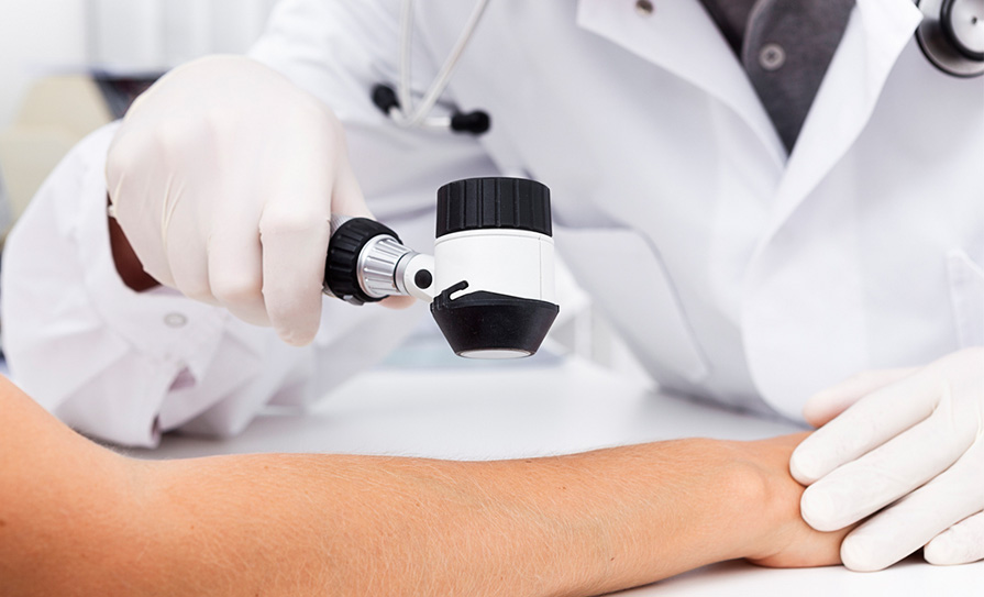
Dr Caroline Burke relays the guidance and advice from the experts Dr Chin Whybrew, Ms Julie Van Onselen, and Dr Julian Pearce, who delivered the Basic Dermoscopy Day at the PCDSI 2023 Annual Scientific Conference, which took place in Kilkenny from 3-4 March.
Dermoscopy helps’ particularly in keeping the basic and benign lesions out of secondary care, ie, reducing referral rates and to reassure yourself that if you think something looks like, for example, a simple warty lesion or, eg, a seborrheic keratosis that that is in fact what it is. Naevi should not grow beyond midlife so any concerning moles, particularly in this age and beyond, should be referred.
‘Dermoscopy is fun’ and there were lots of food analogies used to aid the pattern recognition which is built up over time when viewing lesions dermoscopically. Seborrheic keratosis can resemble chocolate muffins, sebaceous hyperplasia can have a resemblance to marshmallows, and actinic keratosis on the ear can show a ‘cornflake sign’! The advice was to always send any lesion resected away for histology.
Tips were given in terms of types of dermatoscopes – ideally one with options for polarised light and without. Lots of different types are on the market and information on brands is available on the www.pcds.org.uk/ website.
For contact fluids, alcohol gel is advised and simple lubricant for broken skin or around the eye. Clingfilm can be applied on the dermatoscope for broken or bleeding skin for infection control reasons. Simple tape can be used to strip off dead skin/scale from a pigmented lesion. Remember to optimise image quality by wiping excess fluid to remove air bubbles. Alcohol wipes are handy if reviewing numerous lesions over a large area, eg, the back. Some lesions will not require contact with the dermatoscope and it will not be possible with convex surfaces eg, nose. Too much pressure with the dermatoscope can cause issues in viewing the blood vessels, eg, in looking
at a basal cell carcinoma (BCC).
Some other handy tips included using (with consent) the patient’s phone for taking the dermoscopic picture and they can then email it through to the practice via Healthmail in order to file on their chart. This helps with data management and security. A handy app is the ‘Snipping tool’ app to reduce the size of the photo file for saving. It is important to take a localising photo (can show surrounding sun damage), a close-up clinical photo, and a dermoscopy photo. This builds up your knowledge when you get a response from dermatology naming the lesion and can be used for educational purposes.
The speakers were full of energy and enthusiasm, telling the audience that Ireland has the highest level of fake tan users per capita, and showing an interesting way of applying basic table salt to pyogenic granuloma in order to reduce their vascularity and bleeding risk.
Suggested books included Diagnostic Dermatology: The Illustrated Guide by Jonathan Bowling, and Dermoscopy:
The Essentials.
It is important to expose as much of the skin as possible and ensure all make-up is removed, and examine with the naked eye first before using the dermatoscope as not everything needs one.
History from the patient is essential in terms of general history (age/skin type/previous skin concern/immunosuppression/family hx/sun exposure) and history in relation to the lesion (duration/solitary or multiple/any change and timescale of same/crusting, bleeding, itch, pain, inflammation?).
It is important when referring to describe the lesion properly (site/size/single or multiple/shape/colour/surface/papule, nodule, plaque or cyst/tender or not/inflamed or not). Also describe the site and www.anatomymapper.com is a useful site for the exact terminology. Beware always of the ‘ugly duckling’ lesion and always consider when viewing a lesion – could this be a melanoma?
The six common keratinising tumours were covered in detail in terms of their dermoscopic features – actinic keratoses; Bowen’s; squamous cell carcinoma (SCC); keratoacanthoma; seborrheic keratosis; and viral warts. Colours and patterns, and defining and worrying features were discussed over the course of the afternoon. For example, haemangiomas are raised from the surface and are benign overgrowths of blood vessels and are often soft and show a ‘grapefruit segmentation’-type pattern under dermoscopy. There will be red/purple lacunae and a pinkish colour with a sharply-demarcated edge and fibrous white stroma. They routinely never have vessels that cross the middle/go over the top and should not have brown or grey colours. If they are thrombosed, they can look terrifying as they may have black present, which is blood.
Dermatofibromas, which are always palpable from the outset, retain their original morphology and have a dimple sign when you squeeze either side where there is subcutaneous fat as they will shrink inwards, and on dermoscopy classically have a central white structureless area, which is paler than surrounding skin and have peripheral pigment clods.
SCC show a triad of white circles, irregular vessels around the periphery, and a central keratin mass/erosion present. These are often tender and have a high risk of metastases.
One should have a high index of suspicion for BCC if there is a linear lesion on the head or neck which has not healed within four weeks. The patient may say “I think I scratched it” and BCCs may occur in skin creases. Infiltrative BCCs on the face may look like a scar and a good tip is to stretch it and you can see it projecting out in different directions. BCCs under dermoscopy often show arborising vessels on a translucent pink background and can have ulcerations and micro-erosions and white strands and blotches. If there is pigmentation on the BCC always refer for urgent review.
With regard to melanocytic lesions, the worrying features include chaos under dermoscopy and a lack of symmetry, eg, of colour, border, pattern, and more than two colours. Normal benign naevi under dermoscopy should fade out towards the periphery and have a regular pattern/network and they generally do not contain streaks, polymorphous vessels, white lines or blue/white structures. In deciding re asymmetry, one might use the tip: “If this lesion was a pizza – would everyone get the same toppings?” – if not this may be suggestive of chaos in the lesion.
Lots of melanomas are picked up incidentally and are not always the lesion the patient comes in expressing concern about. One should be wary of nail melanomas which contain a pigment band that is widest at the proximal end and has irregular pigmentation, sometimes destroying the nail plate. Ask regarding a history of trauma if there is curved edges, jagged lines, and a purple colour. Drug-induced pigmentation is caused by medications, such as amiodarone, and fungal nails start distally.
Other uses of dermoscopy were described, including looking at head lice; patterns on the skin in scabies; seeing if alopecia areata is scarring or non-scarring; and in looking at lichen planus; and, of course, in assessing diamonds for clarity, etc.
Overall, this was an informative and enjoyable session which the audience enjoyed. It was peppered with sociable coffee and lunch breaks, and lots of quizzes and interaction from the audience.
Dr Lorna Wilson shares her highlights from the Advanced Dermoscopy Course and day two of the 2023 PCDSI Annual Scientific Conference.
After a brief introduction by the chair Dr Kashif Ahmed, we welcomed Dr Jonathan Bowling up to discuss acral melanomas. This was a very comprehensive talk on the importance of distinguishing between benign and malignant nail pigmentation through our dermatoscopes. We were reminded that variable bands of parallel pigmentation and nail dystrophy in the solitary nail, without a history of trauma, was suspicious for melanoma. Hutchinson sign was also discussed, whereby the nailfold is involved and this is indicative of melanoma.
This talk was followed by a remote presentation from Prof Aimilios Lallas on ‘White lesions and white structures’. Here, Prof Lallas differentiated between real white and pseudo white structures; the former being caused as a result of fibrosis, epidermal hyperplasia, and keratin deposits in the skin. The take-home message was that white scar-like fibrosis in a pigmented lesion is always suspicious for melanoma and these are seen much more clearly under polarised light. Keratin plugs are common in seborrheic keratosis and can also be seen in SCC. Finally, epidermal hyperplasia is commonly seen in conditions such as psoriasis and lichen planus.
Prof Giuseppe Argenziano gave a very interesting talk on lesions from head to toe. He discussed benign lesions of the face, including actinic keratosis and solar lentigos, and he had a number of beautiful slides explaining their features. He went through some dermascopic images of melanoma and lentigo maligna and explained their features in detail. He also discussed genital lesions and differentiated between melanosis, which is a benign entity, and melanoma in this region.
Prof Andreas Blum discussed collision lesions in detail with the aid of a number of slides to illustrate his points. He discussed how these lesions are more common in males than in females, 50 per cent of cases are found on the trunk, and the mean age for developing these lesions is between 53-to-60 years of age. In the head and neck region we are more likely to see a BCC collision lesion, and in the trunk we are more likely to see a melanocytic collision lesion. These lesions are linked to a history of higher UV exposure in the past, especially in the head and neck area.
Prof Lallas followed this talk up again remotely on lesions in the mucus membranes. Here he described pigmented lesions of the genital area, which complimented Prof Argenziano’s talk from earlier. He also described non-pigmented lesions in the genital area, such as psoriasis, lichen sclerosus, Zoon balanitis, fungal infections, and Erythroplasia of Queyrat, which is SCC in situ of the penis.
Dr Jonathan Bowling gave a talk on ‘difficult seborrheic keratosis’, which included inflamed, traumatised, hypopigmented, hyperpigmented and clonal seborrheic keratosis. He also explained the immune interaction in seborrheic keratosis and how this changes the colour of seborrheic keratosis to a grey granular pigment.
Prof Argenziano discussed his top tips for the more experienced dermoscopist, and this was followed up with Prof Blum showing us an array of dermascopic images in his talk on chameleon melanoma, which in essence described the variability of melanomas through pictures.
The evening presentations included excellent talks by Prof Mark Davis on lupus and the skin and oral problems. Dr Eoin Storan also gave very interesting presentations on connective tissue diseases and the skin, as well as systemic diseases and the skin. These were very relevant and well received by all in the audience.
Basic general dermatology for everyday practice
Dr Chris Bower started the morning session off with an overview of nails where he discussed onycholysis, onychomycosis, and pterygium in detail.
This was followed up by Prof Anthony Bewley with his interesting talk on psychodermatology. This presentation described delusional infestation and how to manage this very tricky complaint in general practice.
Dr Colin Long gave us the ‘Ten commandments in dermatology’, which described how to avoid common pitfalls in our day-to-day practice, including not describing all rashes as macular-papular!
Dr Brian Malcolm then joined Dr Bower in a very engaging talk on ‘Interesting cases from Devon’, where they went through a number of slides on different unusual presentations to their practice.
Prof Fergal Moloney gave an excellent talk on how to prevent our patients getting skin cancer. He described the predisposing factors for the development of melanoma and the correlation between deprivation and melanoma. He also explained how Nicotinamide twice-daily reduced the risk of developing BCC by 23 per cent.
Dr David Buckley gave a very comprehensive talk on the management of psoriasis in primary care and held an interactive quiz, the winner of which won a copy of his new textbook.
Ms Sheila Ryan gave a very practical talk on wound care where she described the most appropriate dressings for wounds in all their varying stages.
In the advanced dermatology section, Dr Síona Ní Raghallaigh delivered an excellent talk on managing the four different subtypes of rosacea from topical to systemic treatment. She also discussed the options for treating complications of severe rosacea such as disfiguring rhinophyma.
Dr Jerry Tan gave a very detailed and comprehensive presentation on dealing with acne scars. Dr Tan described the different types of acne scars, from ice pick and boxcar to rolling and saucer scars. He went through chemical peels, micro-needling, laser therapy, and dermal fillers and their role in reducing the appearance of disfiguring acne scars. He also spoke about the use of topical retinoids and oral isotretinoin in aiding in this process.
Prof Anthony Bewley was welcomed back to stage again for a very interesting talk on the effects of acne and rosacea on the psyche, and gave very useful pointers on how to deal with patients in these categories.
Finally, Dr Ní Raghallaigh concluded the evening with a very useful talk on the red spotty face not caused by rosacea or acne. Dr Ní Raghallaigh described the management of seborrheic dermatitis, periorofacial dermatitis, steroid rosacea, and pityriasis folliculorum.
We are all very much looking forward to the next PCDSI conference being held in Galway on 11–13 April 2024. For more information go to www.pcdsi.com.


Leave a Reply
You must be logged in to post a comment.