


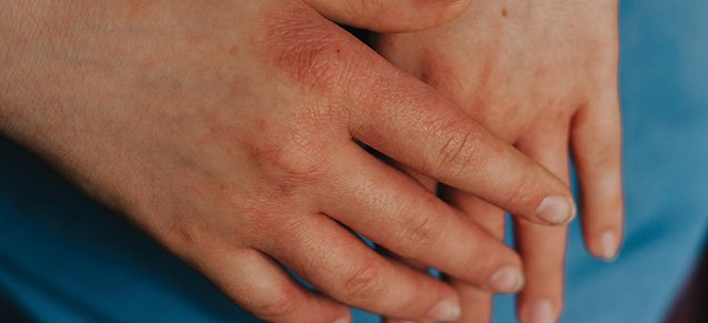
Atopic eczema, also known as atopic dermatitis, is a chronic, inflammatory skin disorder characterised by intense itching, dry skin, and recurrent erythematous lesions, which usually develops in early childhood
The causes of atopic eczema remain unclear, but are multifactorial in nature, involving genetic, socioeconomic, and environmental factors. It can run in families and often develops alongside other atopic conditions such as asthma and allergic rhinitis. The prevalence of atopic eczema in adults is about 1-3 per cent, and 10-20 per cent in children. It affects approximately one-in-five children and one in 12 adults in Ireland. Although it can present at any age, up to 85 per cent of patients are symptomatic before five years of age. The condition is mild in up to 80 per cent of patients; however, it can have a significant negative impact on an individual’s quality-of-life due to itch, sleep deprivation, and social embarrassment.
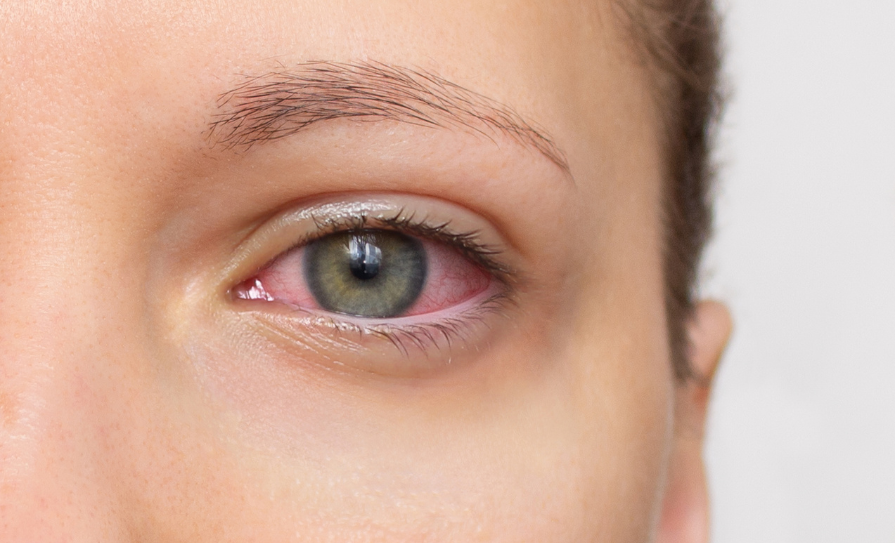
Atopic eczema causes areas of skin to become itchy, dry, cracked, red and sore. It can occur all over the body, but is most common on the hands especially the fingers, insides of the elbows or backs of the knees, and the face and scalp in children. Severity can vary from person to person and although it generally presents as an episodic disease with repeated flare-ups, it can also be continuous. People with mild eczema may only have small areas of dry skin that are occasionally itchy. In more severe cases, atopic eczema can cause widespread red, inflamed skin all over the body and constant itching.
Triggers
Common triggers include irritants such as soaps and detergents; environmental factors such as cold and dry weather, dampness, house dust mites, pet fur, pollen and mold; food allergies such as cows’ milk, eggs, peanuts, soya or wheat; certain materials worn next to the skin, such as wool and synthetic fabrics; hormonal changes; skin infections; and psychological stress.
Pathophysiology
The pathophysiology of atopic eczema is complex, involving elements of barrier dysfunction, alterations in cell-mediated immune responses, IgE-mediated hypersensitivity, and environmental factors. Key components are epidermal barrier dysfunction and cutaneous inflammation due to heightened immune response. The epidermal barrier is made of structural proteins including filaggrin (FLG), whose role is to prevent water loss and penetration of environmental antigens, irritants and microbes. Loss of function mutations in filaggrin have been implicated in severe atopic eczema due to a potential increase in trans-epidermal water loss, pH alterations, and dehydration. However, not all patients with atopic eczema have an FLG mutation and 60 per cent of patients carrying the FLG mutation do not have an atopic disease. The strongest risk factor for atopic eczema is a positive family history for atopic disease. Other genetic changes have also been identified which may alter the skin’s barrier function, resulting in an atopic dermatitis phenotype. The imbalance of Th2 to Th1 cytokines observed in atopic dermatitis can create alterations in the cell-mediated immune responses and can promote IgE mediated hypersensitivity, both of which appear to play a role in the development of atopic eczema. The impact of diet and the role of the environment and chemicals such as airborne formaldehyde, harsh detergents, fragrances, and preservatives must be considered. Use of harsh alkaline detergents in skin care products can alter the skin’s pH causing changes in enzyme activity and triggering inflammation. Environmental pollutants can trigger responses from both the innate and adaptive immune pathways.
Diagnosis
Diagnosis of atopic eczema is mainly based on the patient’s history, clinical findings and exclusion of other dermatological conditions. History includes questions about the onset, pattern and severity of the atopic eczema, potential trigger factors such as irritants, skin infections, contact allergens, food and inhalant allergens, the response to previous and current treatments, the impact of the condition on the patient, and personal/family history of atopic diseases. The diagnosis of atopic eczema relies on the assessment of clinical features because there is no laboratory marker or definitive test available to diagnose the condition. Elevated IgE levels may be seen in up to 80 per cent of patients with atopic eczema, but it is not a specific test for atopic eczema and is not routinely indicated.
No standardised methods to diagnose atopic eczema were available until Hanifin and Rajka developed diagnostic criteria in 1980. Since then, the original criteria have been modified many times and numerous experts have developed different criteria suitable for their own environment, and varying with age. Efforts to develop practical clinical criteria have proven difficult, and those available are not suitable for all geographic areas and age groups. The lack of a good biomarker for diagnosing the condition is considered a major obstacle to the study of atopic eczema. The UK Working Party diagnostic criteria are among those most widely used. However, while the validity and reliability of these criteria have been assessed, they have not been tested extensively in non-white ethnic groups in the UK.
The UK Working Party diagnostic criteria state that patients must have a history of itchy skin and include at least three of the following:
Visible flexural dermatitis involving the skin creases, such as the bends of the elbows or behind the knees (or visible dermatitis on the cheeks and/or extensor areas in children ≤ 18 months).
Personal history of flexural dermatitis (or dermatitis on the cheeks and/or extensor areas in children ≤18 months).
Personal history of dry skin in the last 12 months.
Personal history of asthma or allergic rhinitis (or history of atopic disease in a first-degree relative if the patient is <four years).
Onset of signs and symptoms <two years (this criterion should not be used in children <four years).
A punch skin biopsy may be necessary for patients with atypical presentations to rule out other skin conditions that resemble atopic eczema. These conditions include other inflammatory dermatoses such as seborrheic dermatitis, psoriasis, allergic or irritant contact dermatitis, and pityriasis lichenoides; primary ichthyosis, infestations such as scabies, fungal infections, human immunodeficiency virus (HIV), malignancies, and metabolic disorder.
Atopic eczema should be differentiated from other common dermatological conditions. Allergic contact dermatitis is a differential diagnosis. These two conditions can be challenging to distinguish and both conditions can coexist. With a history or examination suggestive of allergic contact dermatitis, patch testing that can detect delayed hypersensitivity to common allergens should be considered.
Atypical presentations of atopic eczema require further evaluation; for example, adult onset eczematous lesions, which are poorly responsive to topical treatment to exclude cutaneous T-cell lymphoma, and infants with severe extensive eczematous lesions associated with recurrent infections.
Complications
People with atopic eczema and its associated epidermal barrier dysregulation are at risk of secondary infection. Staphylococcus aureus colonises up to 90 per cent of atopic eczema patients compared with 5 per cent of healthy individuals leading to infection and impetiginisation of lesions. Signs and symptoms of secondary bacterial infection include weeping and crusting of lesions, pustules, failing to respond to therapy, rapidly worsening atopic eczema, and rarely fever and malaise. Eczema herpeticum is a severe skin infection with herpes simplex virus that occurs in up to 3 per cent of atopic eczema patients, particularly in severely affected individuals. Children and adults with atopic eczema also have an increased susceptibility to molluscum contagiosum infection. Fungal infections are common in patients with AD. In particular, the yeast M.furfur commonly affects the head neck and trunk with red, hypopigmented or hyperpigmented patches that may have a fine surface scale.
Treatment
While there are a variety of treatments that can help to ease the symptoms, there is no cure for atopic eczema. Treatment and management should be individualised according to each patient’s needs. In addition to pharmacological therapy, cutaneous hydration, identification and elimination of aggravating factors, and relief of pruritus are standard. The treatment of atopic eczema can be demanding, resulting in poor adherence to therapy. Patient education and inclusion in decision-making processes is crucial in providing effective management of the condition, ensuring patient understanding, compliance with treatments and recognition of worsening symptoms. The complexities of the fluctuating condition means that individuals with atopic eczema need direction and support to understand why they need to carry out the recommended ongoing treatments, especially when the skin condition looks normal.
The main treatments for eczema are emollients used every day to stop the skin becoming dry and topical corticosteroid creams and ointments used to reduce swelling and redness during flare-ups. Other treatments include topical calcineurin inhibitors such as topical pimecrolimus or tacrolimus used on sensitive skins that are not responding to simpler treatments. They are particularly effective when used on the faces of children since they seem less likely to produce atrophy.
Additional treatments used to reduce specific symptoms include appropriate medications to treat bacterial, viral or fungal infections should they occur. Antihistamines although not used routinely in atopic eczema, may be used for patients with pruritus causing disturbed sleep.
Emollients remain the mainstay treatment of atopic eczema management and should be used even when the condition is under control. They help to restore the epidermal barrier function by forming an occlusive layer, reduce trans-epidermal water loss and reduce exposure to bacterial and sensitising antigens. Emollients improve the symptoms of itch and pain, reduce the number of flares and reduce the amounts of topical corticosteroids needed to control atopic eczema. Unperfumed emollients should be applied two-to-three-times daily and after bathing to prevent skin drying.
Emollients are available in different formulations including ointments, creams and lotions. Creams are best applied on large and subacute areas, however, ointments are richer in lipids, provide more lubrication and occlusion, and are useful for the treatment of dry and lichenified areas. Lotions have a higher water content and are used to cool or dry strongly inflamed or oozing lesions. Aqueous cream is no longer recommended as an emollient, as it may cause skin irritation including burning, stinging, itching and redness. Emollients should be used in large quantities, up to 250-500g per week.
Topical corticosteroids are effective in treating inflammation and pruritus associated with acute atopic eczema and are recommended as first-line topical therapy of acute flares. The most preferable topical corticosteroids are those that are formulated with low systemic bioavailability and a therapeutic index matched to the area of the involved skin, particularly for infants and young children with widespread involvement. When selecting the potency of the topical corticosteroids, the patient’s age, disease severity, and thickness of the involved skin region/relative absorption should be considered. Topical corticosteroids are classified in terms of strength from mild to very potent. The potency of a topical corticosteroid preparation depends on the formulation as well as the corticosteroid. The inclusion of urea or salicylic acid increases the penetration through the skin.
The least potent but effective topical corticosteroids should be applied once or twice daily. Strength is tailored to the severity of the condition and may vary according to the area of the body being treated. There is evidence to suggest that once daily application of some potent topical corticosteroids may be as effective as twice daily. High-potency topical corticosteroids should not be routinely used on thin skin such as the face, body folds, and groin because of the risk of cutaneous atrophy. Low-potency topical corticosteroids are preferred on the face, areas of thinner skin and in children, however, during acute severe flares, the use of short-term higher-potency topical corticosteroids may be appropriate to control symptoms. Potent and very potent topical corticosteroids should not be used in infants less than 12 months old and very potent topical corticosteroids should not be used in children without specialist dermatological advice. Adverse effects include purpura, telangiectasia, skin atrophy, hypertrichosis, folliculitis, and acne. Most adverse effects will resolve after discontinuing use, but this may take months.
Topical calcineurin inhibitors are anti-inflammatory agents that inhibit calcineurin-dependent T-cell activation. They are used as a second-line treatment for short-term and intermittent care. Tacrolimus 0.03 per cent and 0.1 per cent, applied as a thin layer to the affected area are authorised for use in individuals over 16-years-of-age and tacrolimus 0.03 per cent for children over two years. Topical calcineurin inhibitors do not cause skin atrophy and are useful especially in delicate skin areas such as the face and the groin area. Adverse effects include localised site reactions, eg, stinging and pruritus. Topical calcineurin inhibitors should be avoided for use in individuals with congenital or acquired immunodeficiency and those treated with immunosuppressants or phototherapy. Patients using topical tacrolimus are advised to use sunscreen and minimise sun exposure.
Wet wrap therapy (WWT) involves the application of a topical corticosteroid and/or emollients, under two layers of cotton bandages and is used for short-term, seven-to-14 days control of refractory flares. WWT using topical corticosteroids increase the risk of systemic adverse effects, and should only be initiated by a doctor trained in the procedure.
Optimisation of topical therapy is required before considering systemic therapies. The majority of patients with atopic eczema respond to topical therapy, however for patients with severe atopic eczema not responding to topical therapy systemic immunosuppressants or phototherapy may be required.
Ciclosporin which suppresses T-cells and IL-2 production is used first-line for severe atopic eczema when systemic pharmacological therapy is required. Adverse effects include infection, renal toxicity, hepatotoxicity, hypertension, hyperlipidaemia, hyperkalaemia, risk of lymphomas and malignancies, hypomagnesaemia, bone marrow suppression and potential drug interactions. Monitoring of the patient’s blood pressure, lipids, renal, and liver function is required.
Dupilumab, a targeted therapy, has been authorised for the treatment of moderate to severe atopic eczema. It is a monoclonal antibody administered subcutaneously, that targets IL-4 and IL-13 and has shown efficacy in clinical trials up to one year for the treatment of moderate to severe atopic eczema. Adverse effects include conjunctivitis, herpes simplex, eosinophilia, headache, and injection site reactions.
The use of systemic corticosteroids are discouraged with atopic eczema due to the side effects. They can be used for short-term treatment for less than three weeks in severe circumstances to interrupt flares and when initiating other systemic therapy.
Phototherapy is a therapeutic option for those patients whose atopic eczema cannot be controlled with topical medications alone and who have extensive body spread. It can work in tandem with topical corticosteroids to treat atopic eczema. Phototherapy requires specialist dermatologist supervision and should not be used in combination with topical calcineurin inhibitors or systemic ciclosporin due to a potentially cumulative increased risk of skin cancer.
In summary
The prognosis and outlook for patients with atopic eczema is generally positive, with most children outgrowing the condition by early adolescence. However, patients with severe, widespread disease and concomitant atopic conditions, such as asthma and allergic rhinitis, are likely to experience poorer outcomes. Optimal management of atopic eczema requires a multifaceted approach aimed at healing and protecting the skin barrier and addressing the complex immuno-pathogenesis of the disease. For optimal disease management, patients and/or their caregivers should be educated about the chronic nature of the condition, the need for continued adherence to proper skin care practices, and the appropriate use and application of topical therapies. Poor treatment outcomes are often related to poor adherence, especially to topical therapies, resulting from irrational fears about adverse effects and insufficient information. A number of biologic agents targeting the immune pathways involved in atopic eczema are under investigation representing promising future therapies for the condition.
At least one-third of the population of Ireland is affected by a skin condition. Between 15-20 per cent of GP consultations specifically relate to skin conditions, representing between 3.5 and 4.7 million GP consultations in Ireland each year.
The Irish Skin Foundation (ISF) operates a telephone and email helpline service, which provides direct, accessible and specialist guidance for people affected by all types of skin conditions in Ireland. All ISF nurses are dermatology clinical nurse specialists working in hospitals in Ireland. The ISF helpline is free and available Monday to Friday, 10am – 4:30pm, on (01) 486-6280. See www.irishskin.ie for more information.
References on request


Leave a Reply
You must be logged in to post a comment.