


Attendees at UCD’s Charles Institute Seminar Series heard a presentation by skin research specialist Prof Birgit Lane on keratin filaments and their relationship to genetic skin diseases.
The Charles Institute, Ireland’s national dermatology research and education centre, hosts a range of guest speakers who cover a variety of topics ranging from skin cancer to psoriasis, among many others. The series, which is sponsored by RELIFE (part of the A.Menarini group), is designed to provide expert advice from a range of distinguished national and international experts in their respective fields and is chaired by Prof Desmond Tobin, Full Professor of Dermatological Science at UCD School of Medicine and Director of the Charles Institute of Dermatology. The seminars are broadcast to attendees with a special interest in dermatology and cutaneous science in other locations, who access the talks remotely via an audio-visual link.

The seminars are held using a hybrid model, combining in-person attendance with interactive online access.
Attendees heard a presentation by Prof Birgit Lane, until recently Chief Scientist at the Skin Research Institute of Singapore, and currently Honorary Senior Fellow at A*STAR and Professor emeritus of the University of Dundee. Birgit has decades of research experience in medical cell biology and served as Executive Director of the Institute of Medical Biology, A*STAR, for 11 years. She was also the initiator and co-Executive Director of the Skin Research Institute in Singapore and is the author of more than 260 publications, as well as holding a range of other distinctions in her field.
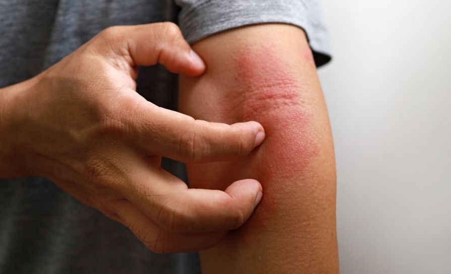
Prof Lane delivered a presentation to the Charles Institute titled ‘What Keratin Filaments Do: Insights from Genetic Skin Diseases’. Keratin proteins form filament networks in skin and other epithelial cells, and following the discoveries of causative roles in epidermolysis bullosa simplex (EBS) and other genetic skin fragility disorders, keratin networks are now known to be essential for tissue resilience, she explained. Analysis of keratinocytes +/- EBS-mimicking mutations in keratins K14 or K5 reveals that EBS-mimetic cells show constitutive stress and many features of a wound-healing activation response, she said. Keratinocyte activation upon stress is likely to be a key evolutionary adaptation of keratinocytes, and keratin changes are an early, persistent aspect of wound-healing, said Prof Lane.
Studies of disease model cells have proven very informative in understanding the regulation and function of keratins in normal cells and have suggested the potential for new approaches to therapy for still-incurable EBS, said Prof Lane.
Keratin mutations
She provided an overview of different types of keratinocytes and intermediate filament genes, with case studies for attendees participating in person and those viewing the presentation online. She described the various different keratinopathies and their different phenotypes, depending on the tissue expression of the mutated keratin. “Epidermal cells with keratin mutations rupture under stress, therefore we conclude that keratins are essential for tissue resilience,” said Prof Lane.
Keratin mutations of EBS also lead to reduced cell junction proteins, said Prof Lane, and oscillating mechanical stress loosens cell-cell junctions, as takes place in the activation of a wound-healing response. In EBS model cells, keratin aggregates begin forming at the cell periphery, and EBS cells are shown to have lower levels of junction proteins, which would also reduce tissue resilience. She pointed out how desmosomes connect keratins from cell-to-cell and may be potential signalling hubs. She also cited research to show that mechanisms of EBS tissue fragility were unlikely to be solely loss of keratin filament resilience, as EBS keratin filaments can withstand extreme stretching: Filament networks break down in response to oscillating stretch, but not to ‘steady’ continuous stretch.
“I think that the keratinocytes activate a wound response when they have been stressed, and in this case the stress is caused by misfolded mutant protein. A number of different mechanisms and types of stress can do that,” Prof Lane told the seminar. “Thinking in an evolutionary way about what keratinocytes do, this is really their main job. If anything happens to the outside layer of the skin, if anything opens-up that outside layer, the keratinocytes must quickly re-seal it, even at the cost of transient loss of resilience. That’s their job; that’s what they have evolved to do, because breaches of that outside barrier are potentially lethal.
“So at any indication that this barrier is in jeopardy, the keratinocytes are ready to migrate towards that wound. It may not initially be a wound, it may be inflammation… but I expect that this is the default behaviour when you stress the keratinocytes,” she continued. “This may be what is happening in EB cells… if you turn down that resilience, the epithelium is going to be fragile. If you live with the skin in that activated state all the time, that is going to be fragile skin. For short periods of wound-healing, that’s fine, but it’s not something you should have to live with,” said Prof Lane.
Cellular stress
“So we think that what we are looking at here is a vicious cycle,” she continued. “Pathogenic keratin mutations lead to aggregate formation; aggregates lead to stress; misfolded protein stress activates keratinocytes; and activation (normally for wound-healing) leads to junctional destabilisation so that the cells can move. But this destabilisation leads to reduced keratin-desmosome connectivity and weakening across the epithelium. Once you start to disconnect and remodel the keratins at the cell’s edge – when the cells are migrating, they need to loosen and remodel that network – once you stimulate that keratin remodelling, the mutant keratins will transiently form more aggregates of misfolded protein. So basically, we think that this is increasing the stress on the cell, and that this increase in stress then has other consequences for the cell.”
That EBS cells are in a constitutive state of stress is born out by multiple lines of experimentation. “Keratinocytes expressing pathogenic keratin mutations show ER stress and a misfolded protein response,” said Prof Lane. “There is also faster response to wounding stress and resistance to apoptosis. In EBS model cells, the stress phenotype is well correlated with keratin aggregates.”
Prof Lane then showed how these disease model cells could be used for drug discovery. She presented their development of a semi-automated cell-based assay for compounds that could specifically reduce stress, and thereby provide new approaches to therapy for EBS. Using optimised EBS-mimicking reporter cells, they developed a computer programme to count aggregate numbers after treatment with a library of phosphorylation-inhibiting compounds. “We ran this assay with a large number of compounds, and we could then identify inhibitors that shut down aggregate formation… and can therefore, we propose, reduce the stress in the cells,” she said. “Compounds like these inhibitors will, we hope, be useful in topical treatment for EBS.”
Phosphorylation is well-known to control many cell signalling processes, as well as keratin assembly and disassembly. Prof Lane explained how differences between different keratin sequences that affect keratin remodelling and bestow different remodelling capacities could affect differentiation. She described how specific phosphorylation sites in keratin proteins may facilitate or inhibit cell division, so that expression of keratins with different phosphorylation capacities could determine cell and tissue function. “The evolution of selective keratin expression helps to determine differentiation and stabilise the tissue. This is a way we can evolve selective keratin function, depending on the fine details of the sequences for phosphorylation.” She suggested that this then helps explain the functional differences between the keratins and the different distribution of keratins in the tissues.
Psoriasis
During a lively Q&A session and clinical discussion following the presentation, Prof Tobin raised a point regarding physiological extremes in conditions, with psoriasis as an example. “In psoriasis, it’s all about proliferation or hyperplasia of keratinocytes, or impoverished differentiation,” he said. “You picked up on the balance between the proliferative vs differentiative [aspects], or even migration vs proliferation – to heal a wound via migration, rather than just ‘packing’ the area with more cells,” he said. “…. In a case like psoriasis, it seems like there should be much more going on in that condition. Do we know anything about the cell fragility in that situation?”
Prof Lane replied: “There are a lot of aspects to consider [in the question] but generally, if you have inflamed skin, the barrier function is almost always compromised, and that’s a more fragile skin. But there are usually a lot more protective cells – there are people who say part of the problem is failure to shed the cells, or part of the reason for the phenotype is failure to shed the cells,” she commented. “We know a lot more about psoriasis these days; you don’t get blistering or detachments, but you get thicker tissue, and perhaps the thicker tissue is partly protective, if there is a compromised barrier.”
Prof Tobin commented: “That is a very good point more generally as we look at biology, in that we may have a condition that guides us towards an expectation of this happening, where the tissue actually compensates by thickening-up as a protective mechanism, which generates a completely different outcome or phenotype than perhaps the initial effect would suggest.”
UCD CHARLES INSTITUTE SEMINAR SERIES.


Leave a Reply
You must be logged in to post a comment.